 Movie
Movie Controller

[English] 日本語
 Yorodumi
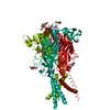
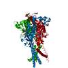
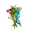
- PDB-5svl: Crystal structure of the ATP-gated human P2X3 ion channel in the ... -
+ Open data

- Basic information
| Entry | Database: PDB / ID: 5svl | |||||||||
|---|---|---|---|---|---|---|---|---|---|---|
| Title | Crystal structure of the ATP-gated human P2X3 ion channel in the ATP-bound, closed (desensitized) state | |||||||||
 Components Components | P2X purinoceptor 3 | |||||||||
 Keywords Keywords | MEMBRANE PROTEIN / Ion Channel / Desensitized State | |||||||||
| Function / homology |  Function and homology information Function and homology informationPlatelet homeostasis / extracellularly ATP-gated monoatomic cation channel activity / purinergic nucleotide receptor activity / Elevation of cytosolic Ca2+ levels / protein homotrimerization / cellular response to ATP / positive regulation of calcium ion transport into cytosol / positive regulation of calcium-mediated signaling / hippocampal mossy fiber to CA3 synapse / modulation of chemical synaptic transmission ...Platelet homeostasis / extracellularly ATP-gated monoatomic cation channel activity / purinergic nucleotide receptor activity / Elevation of cytosolic Ca2+ levels / protein homotrimerization / cellular response to ATP / positive regulation of calcium ion transport into cytosol / positive regulation of calcium-mediated signaling / hippocampal mossy fiber to CA3 synapse / modulation of chemical synaptic transmission / Schaffer collateral - CA1 synapse / calcium ion transmembrane transport / transmembrane transport / signaling receptor complex / postsynapse / axon / signal transduction / ATP binding / metal ion binding / plasma membrane Similarity search - Function | |||||||||
| Biological species |  Homo sapiens (human) Homo sapiens (human) | |||||||||
| Method |  X-RAY DIFFRACTION / SYNCHROTRON / MOLECULAR REPLACEMENT / Resolution: 2.9 Å X-RAY DIFFRACTION / SYNCHROTRON / MOLECULAR REPLACEMENT / Resolution: 2.9 Å | |||||||||
 Authors Authors | Mansoor, S.E. / Lu, W. / Oosterheert, W. / Shekhar, M. / Tajkhorshid, E. / Gouaux, E. | |||||||||
 Citation Citation | Journal: Nature / Year: 2016 Title: X-ray structures define human P2X3 receptor gating cycle and antagonist action. Authors: Mansoor, S.E. / Lu, W. / Oosterheert, W. / Shekhar, M. / Tajkhorshid, E. / Gouaux, E. | |||||||||
| History |
|
- Structure visualization
| Structure viewer | Molecule: MolmilJmol/JSmol |
|---|
- Downloads & links
-Download
| PDBx/mmCIF format | 5svl.cif.gz | 154.5 KB | Display | PDBx/mmCIF format |
|---|---|---|---|---|
| PDB format | pdb5svl.ent.gz | 119.7 KB | Display | PDB format |
| PDBx/mmJSON format | 5svl.json.gz | Tree view | PDBx/mmJSON format | |
| Others |  Other downloads Other downloads |
-Validation report
| Arichive directory | https://data.pdbj.org/pub/pdb/validation_reports/sv/5svlftp://data.pdbj.org/pub/pdb/validation_reports/sv/5svl | HTTPS FTP |
|---|
-Related structure data
| Related structure data |  5svjC  5svkC  5svmC  5svpC  5svqC  5svrC  5svsC  5svtC C: citing same article ( |
|---|---|
| Similar structure data |


-Links
 PDBj
PDBj


- Assembly
| Deposited unit | 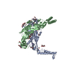
| ||||||||||||
|---|---|---|---|---|---|---|---|---|---|---|---|---|---|
| 1 | 
| ||||||||||||
| 2 | 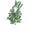
| ||||||||||||
| Unit cell |
| ||||||||||||
| Components on special symmetry positions |
|
-Components
-Protein , 1 types, 2 molecules AB
| #1: Protein | Mass: 40723.828 Da / Num. of mol.: 2 Source method: isolated from a genetically manipulated source Source: (gene. exp.) Homo sapiens (human) / Gene: P2RX3 / Cell line (production host): HEK293 / Production host: Homo sapiens (human) / References: UniProt: P56373 |
|---|
-Sugars , 2 types, 7 molecules 
| #2: Polysaccharide | alpha-D-glucopyranose-(1-4)-alpha-D-glucopyranose / alpha-maltose  Source method: isolated from a genetically manipulated source Details: oligosaccharide / References: alpha-maltose |
|---|---|
| #4: Sugar | ChemComp-NAG /  Type: D-saccharide, beta linking / Mass: 221.208 Da / Num. of mol.: 6 Type: D-saccharide, beta linking / Mass: 221.208 Da / Num. of mol.: 6Source method: isolated from a genetically manipulated source Formula: C8H15NO6 |
-Non-polymers , 6 types, 107 molecules 


| #3: Chemical |  Mass: 507.181 Da / Num. of mol.: 2 Mass: 507.181 Da / Num. of mol.: 2Source method: isolated from a genetically manipulated source Formula: C10H16N5O13P3 / Comment: ATP, energy-carrying molecule*YM #5: Chemical | ChemComp-EDO /  Mass: 62.068 Da / Num. of mol.: 6 / Source method: obtained synthetically / Formula: C2H6O2 Mass: 62.068 Da / Num. of mol.: 6 / Source method: obtained synthetically / Formula: C2H6O2#6: Chemical |  Mass: 59.044 Da / Num. of mol.: 3 / Source method: obtained synthetically / Formula: C2H3O2 Mass: 59.044 Da / Num. of mol.: 3 / Source method: obtained synthetically / Formula: C2H3O2#7: Chemical |  Mass: 22.990 Da / Num. of mol.: 3 / Source method: obtained synthetically / Formula: Na Mass: 22.990 Da / Num. of mol.: 3 / Source method: obtained synthetically / Formula: Na#8: Chemical | ChemComp-TRS / |  Mass: 122.143 Da / Num. of mol.: 1 / Source method: obtained synthetically / Formula: C4H12NO3 / Comment: pH buffer*YM Mass: 122.143 Da / Num. of mol.: 1 / Source method: obtained synthetically / Formula: C4H12NO3 / Comment: pH buffer*YM#9: Water | ChemComp-HOH / | Mass: 18.015 Da / Num. of mol.: 92 / Source method: isolated from a natural source / Formula: H2O |
|---|
-Details
| Has protein modification | Y |
|---|
-Experimental details
-Experiment
| Experiment | Method: X-RAY DIFFRACTION / Number of used crystals: 1 |
|---|
- Sample preparation
| Crystal | Density Matthews: 5.27 Å3/Da / Density % sol: 76.65 % |
|---|---|
| Crystal grow | Temperature: 277.15 K / Method: vapor diffusion, hanging drop Details: 21% PEG 400, 100 mM Tris, pH 8.0, 325 mM Sodium Acetate, 100 mM NaCl |
-Data collection
| Diffraction | Mean temperature: 100 K |
|---|---|
| Diffraction source | Source: SYNCHROTRON / Site: APS  / Beamline: 24-ID-C / Wavelength: 0.979 Å / Beamline: 24-ID-C / Wavelength: 0.979 Å |
| Detector | Type: DECTRIS PILATUS3 S 6M / Detector: PIXEL / Date: Nov 15, 2013 |
| Radiation | Protocol: SINGLE WAVELENGTH / Monochromatic (M) / Laue (L): M / Scattering type: x-ray |
| Radiation wavelength | Wavelength: 0.979 Å / Relative weight: 1 |
| Reflection | Resolution: 2.9→80 Å / Num. obs: 62472 / % possible obs: 97.6 % / Redundancy: 2.84 % / Net I/σ(I): 12.3 |
- Processing
| Software |
| |||||||||||||||||||||||||||||||||||||||||||||||||||||||||||||||||||||||||||||||||||||||||||||||||||||||||||||||||||||||||||||||||||||||||||||||||||||||||||||||||
|---|---|---|---|---|---|---|---|---|---|---|---|---|---|---|---|---|---|---|---|---|---|---|---|---|---|---|---|---|---|---|---|---|---|---|---|---|---|---|---|---|---|---|---|---|---|---|---|---|---|---|---|---|---|---|---|---|---|---|---|---|---|---|---|---|---|---|---|---|---|---|---|---|---|---|---|---|---|---|---|---|---|---|---|---|---|---|---|---|---|---|---|---|---|---|---|---|---|---|---|---|---|---|---|---|---|---|---|---|---|---|---|---|---|---|---|---|---|---|---|---|---|---|---|---|---|---|---|---|---|---|---|---|---|---|---|---|---|---|---|---|---|---|---|---|---|---|---|---|---|---|---|---|---|---|---|---|---|---|---|---|---|---|
| Refinement | Method to determine structure: MOLECULAR REPLACEMENT / Resolution: 2.9→76.983 Å / SU ML: 0.41 / Cross valid method: FREE R-VALUE / σ(F): 1.1 / Phase error: 26.52 / Stereochemistry target values: ML
| |||||||||||||||||||||||||||||||||||||||||||||||||||||||||||||||||||||||||||||||||||||||||||||||||||||||||||||||||||||||||||||||||||||||||||||||||||||||||||||||||
| Solvent computation | Shrinkage radii: 0.9 Å / VDW probe radii: 1.11 Å / Solvent model: FLAT BULK SOLVENT MODEL | |||||||||||||||||||||||||||||||||||||||||||||||||||||||||||||||||||||||||||||||||||||||||||||||||||||||||||||||||||||||||||||||||||||||||||||||||||||||||||||||||
| Refinement step | Cycle: LAST / Resolution: 2.9→76.983 Å
| |||||||||||||||||||||||||||||||||||||||||||||||||||||||||||||||||||||||||||||||||||||||||||||||||||||||||||||||||||||||||||||||||||||||||||||||||||||||||||||||||
| Refine LS restraints |
| |||||||||||||||||||||||||||||||||||||||||||||||||||||||||||||||||||||||||||||||||||||||||||||||||||||||||||||||||||||||||||||||||||||||||||||||||||||||||||||||||
| LS refinement shell |
|
