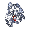
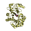
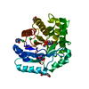
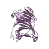
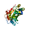
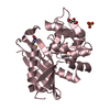
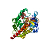
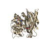
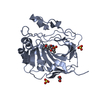
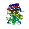
- PDB-4gie: Crystal structure of prostaglandin F synthase from Trypanosoma cr... -
+
Open data
ID or keywords:
Loading...
-
Basic information
Entry
Database: PDB / ID: 4gie
Title
Crystal structure of prostaglandin F synthase from Trypanosoma cruzi bound to NADP
Components
Prostaglandin F synthase
Keywords
OXIDOREDUCTASE / Structural Genomics / NIAID / National Institute of Allergy and Infectious Diseases / Seattle Structural Genomics Center for Infectious Disease / SSGCID / Chagas disease / prostaglandin D2 / trypanosomiasis / eukaryotic pathogen / parasite / NADP+ / NADPH / Rutin / Bimatoprost
Function / homology
Function and homology information
Oxidoreductases; Acting on the CH-OH group of donors; With NAD+ or NADP+ as acceptor / prostaglandin biosynthetic process / oxidoreductase activity, acting on the CH-OH group of donors, NAD or NADP as acceptor / nucleotide binding / cytoplasm Similarity search - Function
Aldo/keto reductase family putative active site signature. / Aldo/keto reductase family signature 1. / NADP-dependent oxidoreductase domain / Aldo/keto reductase family signature 2. / Aldo/keto reductase, conserved site / Aldo-keto reductase / NADP-dependent oxidoreductase domain / Aldo/keto reductase family / NADP-dependent oxidoreductase domain superfamily / TIM Barrel ...Aldo/keto reductase family putative active site signature. / Aldo/keto reductase family signature 1. / NADP-dependent oxidoreductase domain / Aldo/keto reductase family signature 2. / Aldo/keto reductase, conserved site / Aldo-keto reductase / NADP-dependent oxidoreductase domain / Aldo/keto reductase family / NADP-dependent oxidoreductase domain superfamily / TIM Barrel / Alpha-Beta Barrel / Alpha Beta Similarity search - Domain/homology
Mass: 18.015 Da / Num. of mol.: 413 / Source method: isolated from a natural source / Formula: H2O
-
Experimental details
-
Experiment
Experiment
Method: X-RAY DIFFRACTION / Number of used crystals: 1
-
Sample preparation
Crystal
Density Matthews: 2.19 Å3/Da / Density % sol: 43.71 %
Crystal grow
Temperature: 289 K / Method: vapor diffusion, sitting drop / pH: 5.5 Details: TrcrA.00019.a.B1 at 20 mg/mL with 2 mM NADP against Index G6: 0.2 M ammonium acetate, 0.1 M Bis-Tris, pH 5.5, 25% PEG3350, cryoprotectant: 15% ethylene glycol, crystal tracking ID 235869g6, ...Details: TrcrA.00019.a.B1 at 20 mg/mL with 2 mM NADP against Index G6: 0.2 M ammonium acetate, 0.1 M Bis-Tris, pH 5.5, 25% PEG3350, cryoprotectant: 15% ethylene glycol, crystal tracking ID 235869g6, unique puck ID pzu9-6, VAPOR DIFFUSION, SITTING DROP, temperature 289K
-
Data collection
Diffraction
Mean temperature: 100 K
Diffraction source
Source: SYNCHROTRON / Site: ALS / Beamline: 5.0.1 / Wavelength: 0.9774 Å
In the structure databanks used in Yorodumi, some data are registered as the other names, "COVID-19 virus" and "2019-nCoV". Here are the details of the virus and the list of structure data.
Jan 31, 2019. EMDB accession codes are about to change! (news from PDBe EMDB page)
EMDB accession codes are about to change! (news from PDBe EMDB page)
The allocation of 4 digits for EMDB accession codes will soon come to an end. Whilst these codes will remain in use, new EMDB accession codes will include an additional digit and will expand incrementally as the available range of codes is exhausted. The current 4-digit format prefixed with “EMD-” (i.e. EMD-XXXX) will advance to a 5-digit format (i.e. EMD-XXXXX), and so on. It is currently estimated that the 4-digit codes will be depleted around Spring 2019, at which point the 5-digit format will come into force.
The EM Navigator/Yorodumi systems omit the EMD- prefix.
Related info.:Q: What is EMD? / ID/Accession-code notation in Yorodumi/EM Navigator
Yorodumi is a browser for structure data from EMDB, PDB, SASBDB, etc.
This page is also the successor to EM Navigator detail page, and also detail information page/front-end page for Omokage search.
The word "yorodu" (or yorozu) is an old Japanese word meaning "ten thousand". "mi" (miru) is to see.
Related info.:EMDB / PDB / SASBDB / Comparison of 3 databanks / Yorodumi Search / Aug 31, 2016. New EM Navigator & Yorodumi / Yorodumi Papers / Jmol/JSmol / Function and homology information / Changes in new EM Navigator and Yorodumi
 Movie
Movie Controller

 Yorodumi
 Open data
 Basic information
 Components
 Keywords
 Function and homology information
 X-RAY DIFFRACTION /
  Authors
 Citation
 Structure visualization
 Downloads & links
 Other downloads
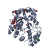


 PDBj
PDBj
 Assembly


 Mass: 59.044 Da / Num. of mol.: 1 / Source method: obtained synthetically / Formula: C2H3O2
 Mass: 743.405 Da / Num. of mol.: 1 / Source method: obtained synthetically / Formula: C21H28N7O17P3
 Mass: 18.015 Da / Num. of mol.: 413 / Source method: isolated from a natural source / Formula: H2O
 Sample preparation
 / Beamline: 5.0.1 / Wavelength: 0.9774 Å
 Processing