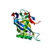
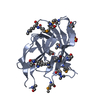
- PDB-4gbs: Crystal structure of a putative lipoprotein (BF2707) from Bactero... -
+
Open data
ID or keywords:
Loading...
-
Basic information
Entry
Database: PDB / ID: 4gbs
Title
Crystal structure of a putative lipoprotein (BF2707) from Bacteroides fragilis NCTC 9343 at 2.75 A resolution
Components
Putative lipoprotein
Keywords
LIPID BINDING PROTEIN / PF14064 family / transport / Structural Genomics / Joint Center for Structural Genomics / JCSG / Protein Structure Initiative / PSI-BIOLOGY / HEME-BINDING PROTEIN
Function / homology
HmuY protein / HmuY protein / Prokaryotic membrane lipoprotein lipid attachment site profile. / Lipoprotein
Function and homology information
Biological species
Bacteroides fragilis (bacteria)
Method
X-RAY DIFFRACTION / SYNCHROTRON / MAD / Resolution: 2.48 Å
Mass: 18.015 Da / Num. of mol.: 72 / Source method: isolated from a natural source / Formula: H2O
Sequence details
1. THE CONSTRUCT (RESIDUES 25-223) WAS EXPRESSED WITH AN N-TERMINAL PURIFICATION TAG ...1. THE CONSTRUCT (RESIDUES 25-223) WAS EXPRESSED WITH AN N-TERMINAL PURIFICATION TAG MGSDKIHHHHHHENLYFQG. THE TAG WAS REMOVED WITH TEV PROTEASE LEAVING ONLY A GLYCINE (0) FOLLOWED BY THE TARGET SEQUENCE. 2. THE PROTEIN WAS REDUCTIVELY METHYLATED PRIOR TO CRYSTALLIZATION.
-
Experimental details
-
Experiment
Experiment
Method: X-RAY DIFFRACTION / Number of used crystals: 1
-
Sample preparation
Crystal
Density Matthews: 3.1 Å3/Da / Density % sol: 60.38 %
Crystal grow
Temperature: 277 K / Method: vapor diffusion, sitting drop / pH: 9 Details: 2.40M ammonium sulfate, 0.1M Bicine pH 9.0, NANODROP, VAPOR DIFFUSION, SITTING DROP, temperature 277K
Resolution: 2.48→46.676 Å / Num. obs: 21701 / % possible obs: 99.6 % / Observed criterion σ(I): -3 / Biso Wilson estimate: 76.09 Å2 / Rmerge F obs: 0.999 / Rmerge(I) obs: 0.072 / Rrim(I) all: 0.078 / Net I/σ(I): 14.65 / Num. measured all: 137544
Reflection shell
Diffraction-ID: 1
Resolution (Å)
Rmerge F obs
Rmerge(I) obs
Mean I/σ(I) obs
Num. measured obs
Num. possible
Num. unique obs
Rrim(I) all
% possible all
2.48-2.57
0.705
1.17
1.7
14273
2138
2133
1.269
99.8
2.57-2.67
0.785
0.775
2.4
13410
2076
2065
0.843
99.5
2.67-2.79
0.87
0.563
3.2
12508
2089
2079
0.617
99.5
2.79-2.94
0.954
0.297
5.7
13972
2178
2176
0.324
99.9
2.94-3.12
0.986
0.18
9.3
14282
2102
2101
0.195
100
3.12-3.36
0.995
0.105
14.4
14460
2162
2157
0.114
99.8
3.36-3.7
0.998
0.067
20.6
14086
2178
2176
0.072
99.9
3.7-4.23
0.998
0.05
24.8
12596
2186
2174
0.055
99.5
4.23-5.31
0.999
0.044
30.8
14381
2222
2215
0.048
99.7
5.31-46.676
0.998
0.05
30
13576
2483
2441
0.055
98.3
-
Phasing
Phasing
Method: MAD
-
Processing
Software
Name
Version
Classification
NB
MolProbity
3beta29
modelbuilding
PDB_EXTRACT
3.1
dataextraction
SHELX
phasing
SHARP
phasing
XSCALE
March15, 2012
datascaling
BUSTER-TNT
2.10.0
refinement
XDS
datareduction
SHELXD
phasing
BUSTER
2.10.0
refinement
Refinement
Method to determine structure: MAD / Resolution: 2.48→46.676 Å / Cor.coef. Fo:Fc: 0.9462 / Cor.coef. Fo:Fc free: 0.9335 / Occupancy max: 1 / Occupancy min: 0.5 / Cross valid method: THROUGHOUT / σ(F): 0 Details: 1. A MET-INHIBITION PROTOCOL WAS USED FOR SELENOMETHIONINE INCORPORATION DURING PROTEIN EXPRESSION. THE OCCUPANCY OF THE SE ATOMS IN THE MSE RESIDUES WAS REDUCED TO 0.75 FOR THE REDUCED ...Details: 1. A MET-INHIBITION PROTOCOL WAS USED FOR SELENOMETHIONINE INCORPORATION DURING PROTEIN EXPRESSION. THE OCCUPANCY OF THE SE ATOMS IN THE MSE RESIDUES WAS REDUCED TO 0.75 FOR THE REDUCED SCATTERING POWER DUE TO PARTIAL S-MET INCORPORATION. 2. ATOM RECORD CONTAINS SUM OF TLS AND RESIDUAL B FACTORS. ANISOU RECORD CONTAINS SUM OF TLS AND RESIDUAL U FACTORS. 3. THE MAD PHASES WERE USED AS RESTRAINTS DURING REFINEMENT. 4. NCS RESTRAINTS WERE APPLIED USING BUSTER'S LSSR RESTRAINT REPRESENTATION (-AUTONCS).5.THE PROTEIN WAS SUBJECTED TO REDUCTIVE METHYLATION PRIOR TO CRYSTALLIZATION. LYSINE 101 APPEARS TO HAVE BEEN PROTECTED FROM REDUCTIVE METHYLATION AND WAS MODELED AS LYSINE. ALL OTHER LYSINES HAVE BEEN MODELED AS N-DIMETHYL-LYSINE (MLY). 6. SULFATE MOLECULES(SO4) FROM THE CRYSTALLIZATION HAVE BEEN MODELED INTO THE STRUCTURE.
In the structure databanks used in Yorodumi, some data are registered as the other names, "COVID-19 virus" and "2019-nCoV". Here are the details of the virus and the list of structure data.
Jan 31, 2019. EMDB accession codes are about to change! (news from PDBe EMDB page)
EMDB accession codes are about to change! (news from PDBe EMDB page)
The allocation of 4 digits for EMDB accession codes will soon come to an end. Whilst these codes will remain in use, new EMDB accession codes will include an additional digit and will expand incrementally as the available range of codes is exhausted. The current 4-digit format prefixed with “EMD-” (i.e. EMD-XXXX) will advance to a 5-digit format (i.e. EMD-XXXXX), and so on. It is currently estimated that the 4-digit codes will be depleted around Spring 2019, at which point the 5-digit format will come into force.
The EM Navigator/Yorodumi systems omit the EMD- prefix.
Related info.:Q: What is EMD? / ID/Accession-code notation in Yorodumi/EM Navigator
Yorodumi is a browser for structure data from EMDB, PDB, SASBDB, etc.
This page is also the successor to EM Navigator detail page, and also detail information page/front-end page for Omokage search.
The word "yorodu" (or yorozu) is an old Japanese word meaning "ten thousand". "mi" (miru) is to see.
Related info.:EMDB / PDB / SASBDB / Comparison of 3 databanks / Yorodumi Search / Aug 31, 2016. New EM Navigator & Yorodumi / Yorodumi Papers / Jmol/JSmol / Function and homology information / Changes in new EM Navigator and Yorodumi
 Movie
Movie Controller

 Yorodumi
 Open data
 Basic information
 Components
 Keywords
 Function and homology information
 Bacteroides fragilis (bacteria)
 X-RAY DIFFRACTION /
  Authors
 Citation
 Structure visualization
 Downloads & links
 Other downloads
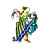

 PDBj
PDBj


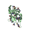
 Assembly
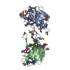

 Mass: 96.063 Da / Num. of mol.: 2 / Source method: obtained synthetically / Formula: SO4
 Mass: 18.015 Da / Num. of mol.: 72 / Source method: isolated from a natural source / Formula: H2O
 Sample preparation
 / Beamline: BL12-2 / Wavelength: 0.9795,0.9184
 Processing