Mass: 18.015 Da / Num. of mol.: 180 / Source method: isolated from a natural source / Formula: H2O
-
Experimental details
-
Experiment
Experiment
Method: X-RAY DIFFRACTION / Number of used crystals: 1
-
Sample preparation
Crystal
Density Matthews: 2.58 Å3/Da / Density % sol: 52.4 %
Crystal grow
Temperature: 298 K / Method: sitting drop vapor diffuction / pH: 7 Details: Protein (10 mM Hepes pH 7.5, 150 mM NaCl, 10% glycerol, 1 mM DTT, 5 mM MgCl); Reservoir (0.1 M Bis-Tris Propane:HCl, 1.8 M Magnesium Sulfate); Cryoprotection (Reservoir, + 20% glycerol), ...Details: Protein (10 mM Hepes pH 7.5, 150 mM NaCl, 10% glycerol, 1 mM DTT, 5 mM MgCl); Reservoir (0.1 M Bis-Tris Propane:HCl, 1.8 M Magnesium Sulfate); Cryoprotection (Reservoir, + 20% glycerol), sitting drop vapor diffuction, temperature 298K
Resolution: 1.7→26.588 Å / Occupancy max: 1 / Occupancy min: 0.06 / FOM work R set: 0.8836 / SU ML: 0.17 / σ(F): 0 / σ(I): 0 / Phase error: 18.81 / Stereochemistry target values: ML Details: THE STRUCTURE WAS REFINED UTILIZING THE STRUCTURE AND GEOMETRIC RESTRAINTS OF BENOZIC ACID, HOWEVER THE EXPERIMENTERS CAN NOT BE CERTAIN OF THE LIGANDS IDENTITY AS BENZOIC ACID WAS NOT A ...Details: THE STRUCTURE WAS REFINED UTILIZING THE STRUCTURE AND GEOMETRIC RESTRAINTS OF BENOZIC ACID, HOWEVER THE EXPERIMENTERS CAN NOT BE CERTAIN OF THE LIGANDS IDENTITY AS BENZOIC ACID WAS NOT A COMPONENT OF THE CRYSTALLIZATION OR PURIFICATION.
Rfactor
Num. reflection
% reflection
Selection details
Rfree
0.2038
1527
5.06 %
RANDOM
Rwork
0.1655
-
-
-
all
0.1673
30180
-
-
obs
0.1673
30180
99.85 %
-
Solvent computation
Shrinkage radii: 0.8 Å / VDW probe radii: 1.1 Å / Solvent model: FLAT BULK SOLVENT MODEL
In the structure databanks used in Yorodumi, some data are registered as the other names, "COVID-19 virus" and "2019-nCoV". Here are the details of the virus and the list of structure data.
Jan 31, 2019. EMDB accession codes are about to change! (news from PDBe EMDB page)
EMDB accession codes are about to change! (news from PDBe EMDB page)
The allocation of 4 digits for EMDB accession codes will soon come to an end. Whilst these codes will remain in use, new EMDB accession codes will include an additional digit and will expand incrementally as the available range of codes is exhausted. The current 4-digit format prefixed with “EMD-” (i.e. EMD-XXXX) will advance to a 5-digit format (i.e. EMD-XXXXX), and so on. It is currently estimated that the 4-digit codes will be depleted around Spring 2019, at which point the 5-digit format will come into force.
The EM Navigator/Yorodumi systems omit the EMD- prefix.
Related info.:Q: What is EMD? / ID/Accession-code notation in Yorodumi/EM Navigator
Yorodumi is a browser for structure data from EMDB, PDB, SASBDB, etc.
This page is also the successor to EM Navigator detail page, and also detail information page/front-end page for Omokage search.
The word "yorodu" (or yorozu) is an old Japanese word meaning "ten thousand". "mi" (miru) is to see.
Related info.:EMDB / PDB / SASBDB / Comparison of 3 databanks / Yorodumi Search / Aug 31, 2016. New EM Navigator & Yorodumi / Yorodumi Papers / Jmol/JSmol / Function and homology information / Changes in new EM Navigator and Yorodumi
 Movie
Movie Controller

 Yorodumi
 Open data
 Basic information
 Components
 Keywords
 Function and homology information
 X-RAY DIFFRACTION /
  Authors
 Citation
 Structure visualization
 Downloads & links
 Other downloads

 PDBj
PDBj


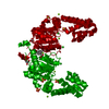
 Assembly
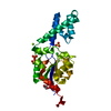


 Mass: 24.305 Da / Num. of mol.: 3 / Source method: obtained synthetically / Formula: Mg
 Mass: 35.453 Da / Num. of mol.: 2 / Source method: obtained synthetically / Formula: Cl
 Mass: 96.063 Da / Num. of mol.: 3 / Source method: obtained synthetically / Formula: SO4
 Mass: 92.094 Da / Num. of mol.: 1 / Source method: obtained synthetically / Formula: C3H8O3
 Sample preparation
 / Beamline: 31-ID / Wavelength: 0.9793 Å
 Processing