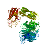
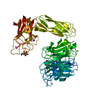
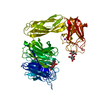
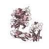
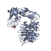
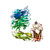
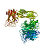
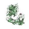
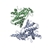
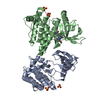
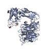
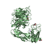
[English] 日本語
Yorodumi
- PDB-1wcq: Mutagenesis of the Nucleophilic Tyrosine in a Bacterial Sialidase... -

+
Open data


ID or keywords:

Loading...

-
Basic information

Entry
Database: PDB / ID: 1wcq
TitleMutagenesis of the Nucleophilic Tyrosine in a Bacterial Sialidase to Phenylalanine.
ComponentsSIALIDASE
KeywordsHYDROLASE / SIALIDASE / MICROMONOSPORA VIRIDIFACIENS / HYDROLASE NEURAMINIDASE / GLYCOSIDASE
Function / homology
Function and homology information


ganglioside catabolic process / oligosaccharide catabolic process / exo-alpha-(2->3)-sialidase activity / exo-alpha-(2->6)-sialidase activity / exo-alpha-(2->8)-sialidase activity / exo-alpha-sialidase / intracellular membrane-bounded organelle / extracellular region / membrane / cytoplasm
Similarity search - Function
Alpha-galactosidase, NEW3 domain / NPCBM-associated, NEW3 domain of alpha-galactosidase / BNR repeat-like domain / Sialidase family / Sialidase / Coagulation factor 5/8 C-terminal domain, discoidin domain / Coagulation factors 5/8 type C domain (FA58C) profile. / F5/8 type C domain / Coagulation factor 5/8 C-terminal domain / Neuraminidase - #10 ...Alpha-galactosidase, NEW3 domain / NPCBM-associated, NEW3 domain of alpha-galactosidase / BNR repeat-like domain / Sialidase family / Sialidase / Coagulation factor 5/8 C-terminal domain, discoidin domain / Coagulation factors 5/8 type C domain (FA58C) profile. / F5/8 type C domain / Coagulation factor 5/8 C-terminal domain / Neuraminidase - #10 / Sialidase superfamily / Galactose-binding domain-like / 6 Propeller / Neuraminidase / Twin arginine translocation (Tat) signal profile. / Twin-arginine translocation pathway, signal sequence / Galactose-binding-like domain superfamily / Immunoglobulin E-set / Jelly Rolls / Immunoglobulins / Immunoglobulin-like fold / Immunoglobulin-like / Sandwich / Mainly Beta
Similarity search - Domain/homology
2-DEOXY-2,3-DEHYDRO-N-ACETYL-NEURAMINIC ACID / Sialidase
Similarity search - Component
Biological speciesMICROMONOSPORA VIRIDIFACIENS (bacteria)
MethodX-RAY DIFFRACTION / SYNCHROTRON / MOLECULAR REPLACEMENT / Resolution: 2.1 Å
AuthorsNewstead, S. / Watson, J.N. / Bennet, A.J. / Taylor, G.
CitationJournal: Chembiochem / Year: 2005
Title: Two Nucleophilic Mutants of the Micromonospora Viridifaciens Sialidase Operate with Retention of Configuration by Two Different Mechanisms.
Authors: Watson, J.N. / Newstead, S. / Narine, A.A. / Taylor, G. / Bennet, A.J.
History
DepositionNov 19, 2004Deposition site: PDBE / Processing site: PDBE
Revision 1.0Oct 12, 2005Provider: repository / Type: Initial release
Revision 1.1Jul 13, 2011Group: Advisory / Version format compliance
Revision 1.2Jul 29, 2020Group: Data collection / Derived calculations / Other
Category: chem_comp / pdbx_database_status ...chem_comp / pdbx_database_status / pdbx_struct_conn_angle / struct_conn / struct_site / struct_site_gen
Item: _chem_comp.type / _pdbx_database_status.status_code_sf ..._chem_comp.type / _pdbx_database_status.status_code_sf / _pdbx_struct_conn_angle.ptnr1_auth_comp_id / _pdbx_struct_conn_angle.ptnr1_auth_seq_id / _pdbx_struct_conn_angle.ptnr1_label_atom_id / _pdbx_struct_conn_angle.ptnr1_label_comp_id / _pdbx_struct_conn_angle.ptnr1_label_seq_id / _pdbx_struct_conn_angle.ptnr3_auth_comp_id / _pdbx_struct_conn_angle.ptnr3_auth_seq_id / _pdbx_struct_conn_angle.ptnr3_label_atom_id / _pdbx_struct_conn_angle.ptnr3_label_comp_id / _pdbx_struct_conn_angle.ptnr3_label_seq_id / _pdbx_struct_conn_angle.value / _struct_conn.pdbx_dist_value / _struct_conn.ptnr1_auth_comp_id / _struct_conn.ptnr1_auth_seq_id / _struct_conn.ptnr1_label_asym_id / _struct_conn.ptnr1_label_atom_id / _struct_conn.ptnr1_label_comp_id / _struct_conn.ptnr1_label_seq_id / _struct_conn.ptnr2_auth_comp_id / _struct_conn.ptnr2_auth_seq_id / _struct_conn.ptnr2_label_asym_id / _struct_conn.ptnr2_label_atom_id / _struct_conn.ptnr2_label_comp_id / _struct_conn.ptnr2_label_seq_id
Description: Carbohydrate remediation / Provider: repository / Type: Remediation
Revision 1.3Dec 13, 2023Group: Data collection / Database references ...Data collection / Database references / Refinement description / Structure summary
Category: chem_comp / chem_comp_atom ...chem_comp / chem_comp_atom / chem_comp_bond / database_2 / pdbx_initial_refinement_model
Item: _chem_comp.pdbx_synonyms / _database_2.pdbx_DOI / _database_2.pdbx_database_accession
Remark 700 SHEET THE SHEET STRUCTURE OF THIS MOLECULE IS BIFURCATED. IN ORDER TO REPRESENT THIS FEATURE IN ... SHEET THE SHEET STRUCTURE OF THIS MOLECULE IS BIFURCATED. IN ORDER TO REPRESENT THIS FEATURE IN THE SHEET RECORDS BELOW, TWO SHEETS ARE DEFINED.

-
Structure visualization

Structure viewerMolecule:
MolmilJmol/JSmol

Downloads & links

-
Assembly

Deposited unit
A: SIALIDASE
B: SIALIDASE
C: SIALIDASE
hetero molecules


Theoretical massNumber of molelcules
Total (without water)194,43516
Polymers192,8483
Non-polymers1,58713
Water19,4201078
1
A: SIALIDASE
hetero molecules


Theoretical massNumber of molelcules
Total (without water)64,7815
Polymers64,2831
Non-polymers4984
Water181
TypeNameSymmetry operationNumber
identity operation1_555x,y,z1
MethodPQS
2
B: SIALIDASE
hetero molecules


Theoretical massNumber of molelcules
Total (without water)64,8736
Polymers64,2831
Non-polymers5915
Water181
TypeNameSymmetry operationNumber
identity operation1_555x,y,z1
MethodPQS
3
C: SIALIDASE
hetero molecules


Theoretical massNumber of molelcules
Total (without water)64,7815
Polymers64,2831
Non-polymers4984
Water181
TypeNameSymmetry operationNumber
identity operation1_555x,y,z1
MethodPQS
Unit cell
Length a, b, c (Å)143.258, 143.258, 160.250
Angle α, β, γ (deg.)90.00, 90.00, 120.00
Int Tables number154
Space group name H-MP3221
Components on special symmetry positions
IDModelComponents
11C-2019-

HOH

-
Components

#1: Protein SIALIDASE / NEURAMINIDASE


Mass: 64282.629 Da / Num. of mol.: 3 / Fragment: SIALIDASE, RESIDUES 47-647 / Mutation: YES
Source method: isolated from a genetically manipulated source
Source: (gene. exp.) MICROMONOSPORA VIRIDIFACIENS (bacteria)
Plasmid: PET28A / Production host: ESCHERICHIA COLI (E. coli) / Strain (production host): BL21(DE3) / References: UniProt: Q02834, exo-alpha-sialidase
#2: Chemical ChemComp-NA / SODIUM ION


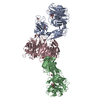
Mass: 22.990 Da / Num. of mol.: 3 / Source method: obtained synthetically / Formula: Na
#3: Chemical
ChemComp-GOL / GLYCEROL / GLYCERIN / PROPANE-1,2,3-TRIOL


Mass: 92.094 Da / Num. of mol.: 7 / Source method: obtained synthetically / Formula: C3H8O3
#4: Sugar ChemComp-DAN / 2-DEOXY-2,3-DEHYDRO-N-ACETYL-NEURAMINIC ACID / Neu5Ac2en


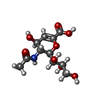
Type: D-saccharide / Mass: 291.255 Da / Num. of mol.: 3 / Source method: obtained synthetically / Formula: C11H17NO8
#5: Water ChemComp-HOH / water


Mass: 18.015 Da / Num. of mol.: 1078 / Source method: isolated from a natural source / Formula: H2O
Compound detailsENGINEERED RESIDUE IN CHAIN A, B, C: TYR 370 TO PHE CATALYTIC ACTIVITY: HYDROLYSIS OF ALPHA-(2->3)- ...ENGINEERED RESIDUE IN CHAIN A, B, C: TYR 370 TO PHE CATALYTIC ACTIVITY: HYDROLYSIS OF ALPHA-(2->3)-, ALPHA-(2->6)-, ALPHA-(2->8)-GLYCOSIDIC LINKAGES OF TERMINAL SIALIC RESIDUES IN OLIGOSACCHARIDES, GLYCOPROTEINS, GLYCOLIPIDS, COLOMINIC ACID AND SYNTHETIC SUBSTRATES.

-
Experimental details

-
Experiment

ExperimentMethod: X-RAY DIFFRACTION / Number of used crystals: 1

-
Sample preparation

CrystalDensity Matthews: 2.3 Å3/Da / Density % sol: 46.7 %
Crystal growpH: 5 / Details: 16 % PEG 3350, 0.2 M AMMONIUM CITRATE, pH 5.00

-
Data collection

DiffractionMean temperature: 100 K
Diffraction sourceSource: SYNCHROTRON / Site: ESRF / Beamline: ID14-2 / Wavelength: 0.933
DetectorType: ADSC CCD / Detector: CCD / Date: Sep 22, 2004 / Details: TOROIDAL MIRROR
RadiationMonochromator: DIAMOND (111), GE(220) / Protocol: SINGLE WAVELENGTH / Monochromatic (M) / Laue (L): M / Scattering type: x-ray
Radiation wavelengthWavelength: 0.933 Å / Relative weight: 1
ReflectionResolution: 2.1→57.83 Å / Num. obs: 110911 / % possible obs: 100 % / Observed criterion σ(I): 6 / Redundancy: 7.2 % / Rmerge(I) obs: 0.07 / Net I/σ(I): 7.4
Reflection shellResolution: 2.1→2.21 Å / Redundancy: 7 % / Rmerge(I) obs: 0.47 / Mean I/σ(I) obs: 1.5 / % possible all: 100

-
Processing

Software
NameVersionClassification
REFMAC5.2.0005refinement
MOSFLMdata reduction
SCALAdata scaling
AMoREphasing
RefinementMethod to determine structure: MOLECULAR REPLACEMENT
Starting model: PDB ENTRY 1EUU
Resolution: 2.1→124.03 Å / Cor.coef. Fo:Fc: 0.958 / Cor.coef. Fo:Fc free: 0.928 / SU B: 9.672 / SU ML: 0.138 / TLS residual ADP flag: LIKELY RESIDUAL / Cross valid method: THROUGHOUT / ESU R: 0.212 / ESU R Free: 0.191 / Stereochemistry target values: MAXIMUM LIKELIHOOD / Details: HYDROGENS HAVE BEEN ADDED IN THE RIDING POSITIONS.
RfactorNum. reflection% reflectionSelection details
Rfree0.238 5544 5 %RANDOM
Rwork0.175 ---
obs0.178 105259 100 %-
Solvent computationIon probe radii: 0.8 Å / Shrinkage radii: 0.8 Å / VDW probe radii: 1.2 Å / Solvent model: MASK
Displacement parametersBiso mean: 29.34 Å2
Baniso -1Baniso -2Baniso -3
1--0.81 Å2-0.4 Å20 Å2
2---0.81 Å20 Å2
3---1.21 Å2
Refinement stepCycle: LAST / Resolution: 2.1→124.03 Å
ProteinNucleic acidLigandSolventTotal
Num. atoms13589 0 105 1078 14772
Refine LS restraints
Refine-IDTypeDev idealDev ideal targetNumber
X-RAY DIFFRACTIONr_bond_refined_d0.0210.02114069
X-RAY DIFFRACTIONr_bond_other_d
X-RAY DIFFRACTIONr_angle_refined_deg1.8351.95219207
X-RAY DIFFRACTIONr_angle_other_deg
X-RAY DIFFRACTIONr_dihedral_angle_1_deg7.11451798
X-RAY DIFFRACTIONr_dihedral_angle_2_deg34.50723.266643
X-RAY DIFFRACTIONr_dihedral_angle_3_deg14.261152043
X-RAY DIFFRACTIONr_dihedral_angle_4_deg19.65115131
X-RAY DIFFRACTIONr_chiral_restr0.1240.22122
X-RAY DIFFRACTIONr_gen_planes_refined0.0080.0211049
X-RAY DIFFRACTIONr_gen_planes_other
X-RAY DIFFRACTIONr_nbd_refined0.2060.26223
X-RAY DIFFRACTIONr_nbd_other
X-RAY DIFFRACTIONr_nbtor_refined0.3080.29311
X-RAY DIFFRACTIONr_nbtor_other
X-RAY DIFFRACTIONr_xyhbond_nbd_refined0.1450.21097
X-RAY DIFFRACTIONr_xyhbond_nbd_other
X-RAY DIFFRACTIONr_metal_ion_refined
X-RAY DIFFRACTIONr_metal_ion_other
X-RAY DIFFRACTIONr_symmetry_vdw_refined0.1870.286
X-RAY DIFFRACTIONr_symmetry_vdw_other
X-RAY DIFFRACTIONr_symmetry_hbond_refined0.2230.236
X-RAY DIFFRACTIONr_symmetry_hbond_other
X-RAY DIFFRACTIONr_symmetry_metal_ion_refined
X-RAY DIFFRACTIONr_symmetry_metal_ion_other
X-RAY DIFFRACTIONr_mcbond_it0.9031.58950
X-RAY DIFFRACTIONr_mcbond_other
X-RAY DIFFRACTIONr_mcangle_it1.448214427
X-RAY DIFFRACTIONr_mcangle_other
X-RAY DIFFRACTIONr_scbond_it2.59335331
X-RAY DIFFRACTIONr_scbond_other
X-RAY DIFFRACTIONr_scangle_it3.8064.54780
X-RAY DIFFRACTIONr_scangle_other
X-RAY DIFFRACTIONr_long_range_B_refined
X-RAY DIFFRACTIONr_long_range_B_other
X-RAY DIFFRACTIONr_rigid_bond_restr
X-RAY DIFFRACTIONr_sphericity_free
X-RAY DIFFRACTIONr_sphericity_bonded
LS refinement shellResolution: 2.1→2.15 Å / Total num. of bins used: 20 /
RfactorNum. reflection
Rfree0.295 430
Rwork0.209 7713
Refinement TLS params.

Method: refined / Refine-ID: X-RAY DIFFRACTION

IDL112)L122)L132)L222)L232)L332)S11 (Å °)S12 (Å °)S13 (Å °)S21 (Å °)S22 (Å °)S23 (Å °)S31 (Å °)S32 (Å °)S33 (Å °)T112)T122)T132)T222)T232)T332)Origin x (Å)Origin y (Å)Origin z (Å)
11.4046-0.32350.01120.6543-0.26260.56170.08160.0646-0.18530.0124-0.0199-0.0572-0.0146-0.0124-0.0617-0.0777-0.0614-0.0079-0.1067-0.0930.0643-24.82659.37284.872
21.84080.24451.74740.90481.30866.38840.29880.137-0.20060.03850.202-0.18570.32680.5608-0.5008-0.17410.06820.0076-0.1228-0.12210.2595.60547.78968.377
32.48650.5322-0.63122.0841-0.80171.08630.0810.06430.0037-0.05640.11190.15610.1550.007-0.1929-0.1236-0.03640.0178-0.0949-0.10270.0631-14.46458.36745.273
40.6535-0.4412-0.21412.4941-0.18620.51940.02170.1391-0.07340.2022-0.06940.0967-0.127-0.0060.0477-0.05720.00420.03310.0044-0.0427-0.109762.724-27.63941.414
54.4861-0.94151.38811.8552-0.45781.0476-0.0120.27830.08880.1514-0.04160.007-0.30920.16930.05360.0753-0.00680.0721-0.06160.0494-0.125153.7637.64738.464
60.29720.36160.20692.8697-0.51140.8385-0.0246-0.00610.02370.14780.26370.5106-0.1135-0.1289-0.2391-0.05590.13860.1342-0.02490.11920.077226.077-8.71642.769
70.23650.2242-0.03030.9287-0.18740.9509-0.09620.02710.09510.07950.09630.1325-0.1921-0.00640-0.03110.018-0.0418-0.1010.040.0488-0.92422.70427.289
87.87261.2259-4.4630.8041-1.08544.03480.071-0.9053-0.2436-0.1363-0.29430.0237-0.19260.87930.22320.0586-0.045-0.00370.05730.0594-0.152517.04923.24858.086
91.75820.3658-0.5492.07480.15011.84040.0128-0.3252-0.03830.0842-0.0401-0.0226-0.09490.26590.0273-0.0859-0.00780.026-0.01320.0475-0.020237.62534.56933.936
Refinement TLS group
IDRefine-IDRefine TLS-IDAuth asym-IDAuth seq-ID
1X-RAY DIFFRACTION1A47 - 402
2X-RAY DIFFRACTION2A403 - 502
3X-RAY DIFFRACTION3A503 - 647
4X-RAY DIFFRACTION4B47 - 402
5X-RAY DIFFRACTION5B403 - 502
6X-RAY DIFFRACTION6B503 - 647
7X-RAY DIFFRACTION7C49 - 402
8X-RAY DIFFRACTION8C403 - 502
9X-RAY DIFFRACTION9C503 - 647

+
About Yorodumi

-
News

-
Feb 9, 2022. New format data for meta-information of EMDB entries

New format data for meta-information of EMDB entries

  • Version 3 of the EMDB header file is now the official format.
  • The previous official version 1.9 will be removed from the archive.

Related info.:EMDB header

External links:wwPDB to switch to version 3 of the EMDB data model

-
Aug 12, 2020. Covid-19 info

Covid-19 info

URL: https://pdbjlc1.pdbj.org/emnavi/covid19.php

New page: Covid-19 featured information page in EM Navigator.

Related info.:Covid-19 info / Mar 5, 2020. Novel coronavirus structure data

+
Mar 5, 2020. Novel coronavirus structure data

Novel coronavirus structure data

Related info.:Yorodumi Speices / Aug 12, 2020. Covid-19 info

External links:COVID-19 featured content - PDBj / Molecule of the Month (242):Coronavirus Proteases

+
Jan 31, 2019. EMDB accession codes are about to change! (news from PDBe EMDB page)

EMDB accession codes are about to change! (news from PDBe EMDB page)

  • The allocation of 4 digits for EMDB accession codes will soon come to an end. Whilst these codes will remain in use, new EMDB accession codes will include an additional digit and will expand incrementally as the available range of codes is exhausted. The current 4-digit format prefixed with “EMD-” (i.e. EMD-XXXX) will advance to a 5-digit format (i.e. EMD-XXXXX), and so on. It is currently estimated that the 4-digit codes will be depleted around Spring 2019, at which point the 5-digit format will come into force.
  • The EM Navigator/Yorodumi systems omit the EMD- prefix.

Related info.:Q: What is EMD? / ID/Accession-code notation in Yorodumi/EM Navigator

External links:EMDB Accession Codes are Changing Soon! / Contact to PDBj

+
Jul 12, 2017. Major update of PDB

Major update of PDB

  • wwPDB released updated PDB data conforming to the new PDBx/mmCIF dictionary.
  • This is a major update changing the version number from 4 to 5, and with Remediation, in which all the entries are updated.
  • In this update, many items about electron microscopy experimental information are reorganized (e.g. em_software).
  • Now, EM Navigator and Yorodumi are based on the updated data.

External links:wwPDB Remediation / Enriched Model Files Conforming to OneDep Data Standards Now Available in the PDB FTP Archive

-
Yorodumi

Thousand views of thousand structures

  • Yorodumi is a browser for structure data from EMDB, PDB, SASBDB, etc.
  • This page is also the successor to EM Navigator detail page, and also detail information page/front-end page for Omokage search.
  • The word "yorodu" (or yorozu) is an old Japanese word meaning "ten thousand". "mi" (miru) is to see.

Related info.:EMDB / PDB / SASBDB / Comparison of 3 databanks / Yorodumi Search / Aug 31, 2016. New EM Navigator & Yorodumi / Yorodumi Papers / Jmol/JSmol / Function and homology information / Changes in new EM Navigator and Yorodumi

Read more