 Movie
Movie Controller

[English] 日本語
 Yorodumi
- PDB-1w8n: Contribution of the Active Site Aspartic Acid to Catalysis in the... -
+ Open data

- Basic information
| Entry | Database: PDB / ID: 1w8n | ||||||
|---|---|---|---|---|---|---|---|
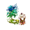
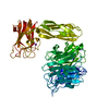
| Title | Contribution of the Active Site Aspartic Acid to Catalysis in the Bacterial Neuraminidase from Micromonospora viridifaciens. | ||||||
 Components Components | BACTERIAL SIALIDASE | ||||||
 Keywords Keywords | HYDROLASE / GLYCOSIDASE / NEURAMINIDASE / BETA- PROPELLER FOLD. | ||||||
| Function / homology |  Function and homology information Function and homology informationganglioside catabolic process / oligosaccharide catabolic process / exo-alpha-sialidase / exo-alpha-sialidase activity / extracellular region / membrane / cytoplasm Similarity search - Function | ||||||
| Biological species |  MICROMONOSPORA VIRIDIFACIENS (bacteria) MICROMONOSPORA VIRIDIFACIENS (bacteria) | ||||||
| Method |  X-RAY DIFFRACTION / MOLECULAR REPLACEMENT / Resolution: 2.1 Å X-RAY DIFFRACTION / MOLECULAR REPLACEMENT / Resolution: 2.1 Å | ||||||
 Authors Authors | Newstead, S. / Watson, J.N. / Dookhun, V. / Bennet, A.J. / Taylor, G. | ||||||
 Citation Citation | Journal: FEBS Lett. / Year: 2004 Title: Contribution of the Active Site Aspartic Acid to Catalysis in the Bacterial Neuraminidase from Micromonospora Viridifaciens Authors: Watson, J.N. / Newstead, S. / Dookhun, V. / Taylor, G. / Bennet, A.J. | ||||||
| History |
| ||||||
| Remark 700 | SHEET THE SHEET STRUCTURE OF THIS MOLECULE IS BIFURCATED. IN ORDER TO REPRESENT THIS FEATURE IN ... SHEET THE SHEET STRUCTURE OF THIS MOLECULE IS BIFURCATED. IN ORDER TO REPRESENT THIS FEATURE IN THE SHEET RECORDS BELOW, TWO SHEETS ARE DEFINED. |
- Structure visualization
| Structure viewer | Molecule: MolmilJmol/JSmol |
|---|
- Downloads & links
-Download
| PDBx/mmCIF format | 1w8n.cif.gz | 140 KB | Display | PDBx/mmCIF format |
|---|---|---|---|---|
| PDB format | pdb1w8n.ent.gz | 107.2 KB | Display | PDB format |
| PDBx/mmJSON format | 1w8n.json.gz | Tree view | PDBx/mmJSON format | |
| Others |  Other downloads Other downloads |
-Validation report
| Arichive directory | https://data.pdbj.org/pub/pdb/validation_reports/w8/1w8nftp://data.pdbj.org/pub/pdb/validation_reports/w8/1w8n | HTTPS FTP |
|---|
-Related structure data
| Related structure data | 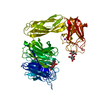 1w8oC 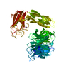 1eutS S: Starting model for refinement C: citing same article ( |
|---|---|
| Similar structure data |


-Links
 PDBj
PDBj


- Assembly
| Deposited unit | 
| ||||||||
|---|---|---|---|---|---|---|---|---|---|
| 1 |
| ||||||||
| Unit cell |
|
-Components
| #1: Protein | Mass: 64240.598 Da / Num. of mol.: 1 / Fragment: RESIDUES 47-647 / Mutation: YES Source method: isolated from a genetically manipulated source Source: (gene. exp.) MICROMONOSPORA VIRIDIFACIENS (bacteria)Plasmid: PET28A / Production host: | ||
|---|---|---|---|
| #2: Sugar | ChemComp-GAL /   Type: D-saccharide, beta linking / Mass: 180.156 Da / Num. of mol.: 1 Type: D-saccharide, beta linking / Mass: 180.156 Da / Num. of mol.: 1Source method: isolated from a genetically manipulated source Formula: C6H12O6 | ||
| #3: Sugar | ChemComp-DAN /   Type: D-saccharide / Mass: 291.255 Da / Num. of mol.: 1 / Source method: obtained synthetically / Formula: C11H17NO8 Type: D-saccharide / Mass: 291.255 Da / Num. of mol.: 1 / Source method: obtained synthetically / Formula: C11H17NO8 | ||
| #4: Chemical | ChemComp-NA /   Mass: 22.990 Da / Num. of mol.: 1 / Source method: obtained synthetically / Formula: Na Mass: 22.990 Da / Num. of mol.: 1 / Source method: obtained synthetically / Formula: Na | ||
| #5: Water | ChemComp-HOH /  Mass: 18.015 Da / Num. of mol.: 516 / Source method: isolated from a natural source / Formula: H2O Mass: 18.015 Da / Num. of mol.: 516 / Source method: isolated from a natural source / Formula: H2O | ||
| Compound details | ENGINEERED| Has protein modification | Y | |
-Experimental details
-Experiment
| Experiment | Method: X-RAY DIFFRACTION / Number of used crystals: 1 |
|---|
- Sample preparation
| Crystal | Density Matthews: 2.8 Å3/Da / Density % sol: 54.9 % |
|---|---|
| Crystal grow | Details: 16 % PEG 3350, 0.2M AMMONIUM CITRATE. |
-Data collection
| Diffraction | Mean temperature: 100 K |
|---|---|
| Diffraction source | Source: ROTATING ANODE / Type: RIGAKU MICROMAX-007 / Wavelength: 1.5418 |
| Detector | Type: RIGAKU-MSC / Detector: IMAGE PLATE / Date: May 6, 2003 / Details: OSMIC MIRRORS |
| Radiation | Protocol: SINGLE WAVELENGTH / Monochromatic (M) / Laue (L): M / Scattering type: x-ray |
| Radiation wavelength | Wavelength: 1.5418 Å / Relative weight: 1 |
| Reflection | Resolution: 2.1→19.92 Å / Num. obs: 44697 / % possible obs: 89 % / Observed criterion σ(I): 6 / Redundancy: 5.6 % / Rmerge(I) obs: 0.07 / Net I/σ(I): 6.9 |
- Processing
| Software |
| ||||||||||||||||||||||||||||||||||||||||||||||||||||||||||||||||||||||||||||||||||||||||||||||||||||||||||||||||||||||||||||||||||||||||||||||||||||||||||||||||||||||||||||||||||||||
|---|---|---|---|---|---|---|---|---|---|---|---|---|---|---|---|---|---|---|---|---|---|---|---|---|---|---|---|---|---|---|---|---|---|---|---|---|---|---|---|---|---|---|---|---|---|---|---|---|---|---|---|---|---|---|---|---|---|---|---|---|---|---|---|---|---|---|---|---|---|---|---|---|---|---|---|---|---|---|---|---|---|---|---|---|---|---|---|---|---|---|---|---|---|---|---|---|---|---|---|---|---|---|---|---|---|---|---|---|---|---|---|---|---|---|---|---|---|---|---|---|---|---|---|---|---|---|---|---|---|---|---|---|---|---|---|---|---|---|---|---|---|---|---|---|---|---|---|---|---|---|---|---|---|---|---|---|---|---|---|---|---|---|---|---|---|---|---|---|---|---|---|---|---|---|---|---|---|---|---|---|---|---|---|
| Refinement | Method to determine structure: MOLECULAR REPLACEMENT Starting model: PDB ENTRY 1EUT Resolution: 2.1→87.71 Å / Cor.coef. Fo:Fc: 0.961 / Cor.coef. Fo:Fc free: 0.935 / SU B: 4.467 / SU ML: 0.119 / TLS residual ADP flag: LIKELY RESIDUAL / Cross valid method: THROUGHOUT / ESU R: 0.202 / ESU R Free: 0.18 / Stereochemistry target values: MAXIMUM LIKELIHOOD / Details: HYDROGENS HAVE BEEN ADDED IN THE RIDING POSITIONS.
| ||||||||||||||||||||||||||||||||||||||||||||||||||||||||||||||||||||||||||||||||||||||||||||||||||||||||||||||||||||||||||||||||||||||||||||||||||||||||||||||||||||||||||||||||||||||
| Solvent computation | Ion probe radii: 0.8 Å / Shrinkage radii: 0.8 Å / VDW probe radii: 1.4 Å / Solvent model: BABINET MODEL WITH MASK | ||||||||||||||||||||||||||||||||||||||||||||||||||||||||||||||||||||||||||||||||||||||||||||||||||||||||||||||||||||||||||||||||||||||||||||||||||||||||||||||||||||||||||||||||||||||
| Displacement parameters | Biso mean: 23.41 Å2
| ||||||||||||||||||||||||||||||||||||||||||||||||||||||||||||||||||||||||||||||||||||||||||||||||||||||||||||||||||||||||||||||||||||||||||||||||||||||||||||||||||||||||||||||||||||||
| Refinement step | Cycle: LAST / Resolution: 2.1→87.71 Å
| ||||||||||||||||||||||||||||||||||||||||||||||||||||||||||||||||||||||||||||||||||||||||||||||||||||||||||||||||||||||||||||||||||||||||||||||||||||||||||||||||||||||||||||||||||||||
| Refine LS restraints |
|