 Movie
Movie Controller

[English] 日本語
 Yorodumi
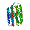
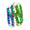
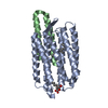
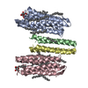
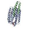
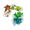
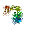
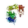
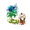
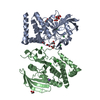
- PDB-1h2s: Molecular basis of transmenbrane signalling by sensory rhodopsin ... -
+ Open data

- Basic information
| Entry | Database: PDB / ID: 1h2s | ||||||
|---|---|---|---|---|---|---|---|
| Title | Molecular basis of transmenbrane signalling by sensory rhodopsin II-transducer complex | ||||||
 Components Components |
| ||||||
 Keywords Keywords | MEMBRANE PROTEIN / MENBRANE PROTEIN COMPLEX / SIGNAL TRANSDUCTION | ||||||
| Function / homology |  Function and homology information Function and homology informationphotoreceptor activity / phototransduction / monoatomic ion channel activity / chemotaxis / transmembrane signaling receptor activity / signal transduction / identical protein binding / plasma membrane Similarity search - Function | ||||||
| Biological species |  NATRONOMONAS PHARAONIS (archaea) NATRONOMONAS PHARAONIS (archaea) | ||||||
| Method |  X-RAY DIFFRACTION / SYNCHROTRON / MOLECULAR REPLACEMENT / Resolution: 1.93 Å X-RAY DIFFRACTION / SYNCHROTRON / MOLECULAR REPLACEMENT / Resolution: 1.93 Å | ||||||
 Authors Authors | Gordeliy, V.I. / Labahn, J. / Moukhametzianov, R. / Efremov, R. / Granzin, J. / Schlesinger, R. / Bueldt, G. / Savopol, T. / Scheidig, A. / Klare, J.P. / Engelhard, M. | ||||||
 Citation Citation | Journal: Nature / Year: 2002 Title: Molecular Basis of Transmembrane Signalling by Sensory Rhodopsin II-Transducer Complex Authors: Gordeliy, V.I. / Labahn, J. / Moukhametzianov, R. / Efremov, R. / Granzin, J. / Schlesinger, R. / Bueldt, G. / Savopol, T. / Scheidig, A. / Klare, J.P. / Engelhard, M. | ||||||
| History |
|
- Structure visualization
| Structure viewer | Molecule: MolmilJmol/JSmol |
|---|
- Downloads & links
-Download
| PDBx/mmCIF format | 1h2s.cif.gz | 66.8 KB | Display | PDBx/mmCIF format |
|---|---|---|---|---|
| PDB format | pdb1h2s.ent.gz | 48.4 KB | Display | PDB format |
| PDBx/mmJSON format | 1h2s.json.gz | Tree view | PDBx/mmJSON format | |
| Others |  Other downloads Other downloads |
-Validation report
| Arichive directory | https://data.pdbj.org/pub/pdb/validation_reports/h2/1h2sftp://data.pdbj.org/pub/pdb/validation_reports/h2/1h2s | HTTPS FTP |
|---|
-Related structure data
| Related structure data | 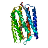 1jgjS S: Starting model for refinement |
|---|---|
| Similar structure data |

-Links
 PDBj
PDBj


- Assembly
| Deposited unit | 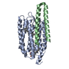
| ||||||||
|---|---|---|---|---|---|---|---|---|---|
| 1 | 
| ||||||||
| Unit cell |
|
-Components
| #1: Protein | Mass: 24085.465 Da / Num. of mol.: 1 / Fragment: RESIDUES 1-225 Source method: isolated from a genetically manipulated source Details: HIS-TAG / Source: (gene. exp.) NATRONOMONAS PHARAONIS (archaea) / Plasmid: PET27BMOD / Production host:  |
|---|---|
| #2: Protein | Mass: 5714.688 Da / Num. of mol.: 1 / Fragment: RESIDUES 23-82 Source method: isolated from a genetically manipulated source Details: HIS-TAG / Source: (gene. exp.) NATRONOMONAS PHARAONIS (archaea) / Plasmid: PET27BMOD / Production host: |
| #3: Sugar | ChemComp-BOG /   Type: D-saccharide / Mass: 292.369 Da / Num. of mol.: 1 Type: D-saccharide / Mass: 292.369 Da / Num. of mol.: 1Source method: isolated from a genetically manipulated source Formula: C14H28O6 / Comment: detergent*YM |
| #4: Chemical | ChemComp-RET /   Mass: 284.436 Da / Num. of mol.: 1 / Source method: obtained synthetically / Formula: C20H28O Mass: 284.436 Da / Num. of mol.: 1 / Source method: obtained synthetically / Formula: C20H28O |
| #5: Water | ChemComp-HOH /  Mass: 18.015 Da / Num. of mol.: 40 / Source method: isolated from a natural source / Formula: H2O Mass: 18.015 Da / Num. of mol.: 40 / Source method: isolated from a natural source / Formula: H2O |
| Compound details | SENSORY RHODOPSIN II INVOLVED IN CONTROL OF PHOTOTAXIS. SEEMS TO ACTIVATE A METHYL-ACCEPTING ...SENSORY RHODOPSIN II INVOLVED IN CONTROL OF PHOTOTAXIS |
| Has protein modification | Y |
-Experimental details
-Experiment
| Experiment | Method: X-RAY DIFFRACTION / Number of used crystals: 1 |
|---|
- Sample preparation
| Crystal | Density Matthews: 2.64 Å3/Da / Density % sol: 53.35 % | ||||||||||||||||||||||||||||
|---|---|---|---|---|---|---|---|---|---|---|---|---|---|---|---|---|---|---|---|---|---|---|---|---|---|---|---|---|---|
| Crystal grow | Temperature: 295 K / Method: lipidic cubic phase / pH: 5.1 Details: 150 MM NACL, 25 MM NAKPI 5.1 0.8% B-OCTYLGLUCOSID , MONOVACCENIN (CUBIC PHASE) PRECIPITATED BY 1 M NA/KPI 5.8 AT 22 C, pH 5.10 | ||||||||||||||||||||||||||||
| Crystal grow | *PLUS Temperature: 22 ℃ / Method: unknown | ||||||||||||||||||||||||||||
| Components of the solutions | *PLUS
|
-Data collection
| Diffraction | Mean temperature: 100 K |
|---|---|
| Diffraction source | Source: SYNCHROTRON / Site: ESRF  / Beamline: ID14-1 / Wavelength: 0.934 / Beamline: ID14-1 / Wavelength: 0.934 |
| Detector | Detector: CCD |
| Radiation | Protocol: SINGLE WAVELENGTH / Monochromatic (M) / Laue (L): M / Scattering type: x-ray |
| Radiation wavelength | Wavelength: 0.934 Å / Relative weight: 1 |
| Reflection | Resolution: 1.93→60 Å / Num. obs: 23156 / % possible obs: 94.7 % / Observed criterion σ(I): 0 / Redundancy: 3.5 % / Rmerge(I) obs: 0.062 / Net I/σ(I): 5.5 |
| Reflection shell | *PLUS % possible obs: 77.3 % / Rmerge(I) obs: 0.394 / Mean I/σ(I) obs: 1.7 |
- Processing
| Software |
| ||||||||||||||||||||||||||||||||||||||||||||||||||||||||||||
|---|---|---|---|---|---|---|---|---|---|---|---|---|---|---|---|---|---|---|---|---|---|---|---|---|---|---|---|---|---|---|---|---|---|---|---|---|---|---|---|---|---|---|---|---|---|---|---|---|---|---|---|---|---|---|---|---|---|---|---|---|---|
| Refinement | Method to determine structure: MOLECULAR REPLACEMENT Starting model: PDB ENTRY 1JGJ Resolution: 1.93→60 Å / Data cutoff high absF: 1000 / Isotropic thermal model: RESTRAINED / Cross valid method: THROUGHOUT / σ(F): 0
| ||||||||||||||||||||||||||||||||||||||||||||||||||||||||||||
| Solvent computation | Solvent model: FLAT | ||||||||||||||||||||||||||||||||||||||||||||||||||||||||||||
| Displacement parameters | Biso mean: 32.3815 Å2
| ||||||||||||||||||||||||||||||||||||||||||||||||||||||||||||
| Refine analyze |
| ||||||||||||||||||||||||||||||||||||||||||||||||||||||||||||
| Refinement step | Cycle: LAST / Resolution: 1.93→60 Å
| ||||||||||||||||||||||||||||||||||||||||||||||||||||||||||||
| Refine LS restraints |
| ||||||||||||||||||||||||||||||||||||||||||||||||||||||||||||
| LS refinement shell | Resolution: 1.93→2 Å / Total num. of bins used: 10 /
| ||||||||||||||||||||||||||||||||||||||||||||||||||||||||||||
| Xplor file |
| ||||||||||||||||||||||||||||||||||||||||||||||||||||||||||||
| Refinement | *PLUS % reflection Rfree: 5 % / Rfactor Rfree: 0.258 / Rfactor Rwork: 0.228 | ||||||||||||||||||||||||||||||||||||||||||||||||||||||||||||
| Solvent computation | *PLUS | ||||||||||||||||||||||||||||||||||||||||||||||||||||||||||||
| Displacement parameters | *PLUS | ||||||||||||||||||||||||||||||||||||||||||||||||||||||||||||
| Refine LS restraints | *PLUS
|
