 Movie
Movie Controller

[English] 日本語
 Yorodumi
- PDB-4f57: Fab structure of a neutralizing antibody L1 from an early subtype... -
+ Open data

- Basic information
| Entry | Database: PDB / ID: 4f57 | ||||||
|---|---|---|---|---|---|---|---|
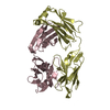
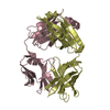
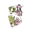
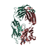
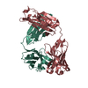
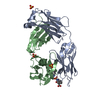
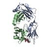
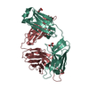
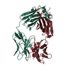
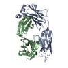
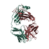
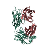
| Title | Fab structure of a neutralizing antibody L1 from an early subtype A HIV-1 infected patient | ||||||
 Components Components |
| ||||||
 Keywords Keywords | IMMUNE SYSTEM / Ig / antibody / gp120 | ||||||
| Function / homology | Immunoglobulins / Immunoglobulin-like / Sandwich / Mainly Beta Function and homology information Function and homology information | ||||||
| Biological species |  Homo sapiens (human) Homo sapiens (human) | ||||||
| Method |  X-RAY DIFFRACTION / SYNCHROTRON / MOLECULAR REPLACEMENT / Resolution: 1.7 Å X-RAY DIFFRACTION / SYNCHROTRON / MOLECULAR REPLACEMENT / Resolution: 1.7 Å | ||||||
 Authors Authors | Pan, R.M. / Kong, X.P. | ||||||
 Citation Citation | Journal: Plos Pathog. / Year: 2013 Title: Viral Escape from Neutralizing Antibodies in Early Subtype A HIV-1 Infection Drives an Increase in Autologous Neutralization Breadth. Authors: Murphy, M.K. / Yue, L. / Pan, R. / Boliar, S. / Sethi, A. / Tian, J. / Pfafferot, K. / Karita, E. / Allen, S.A. / Cormier, E. / Goepfert, P.A. / Borrow, P. / Robinson, J.E. / Gnanakaran, S. ...Authors: Murphy, M.K. / Yue, L. / Pan, R. / Boliar, S. / Sethi, A. / Tian, J. / Pfafferot, K. / Karita, E. / Allen, S.A. / Cormier, E. / Goepfert, P.A. / Borrow, P. / Robinson, J.E. / Gnanakaran, S. / Hunter, E. / Kong, X.P. / Derdeyn, C.A. | ||||||
| History |
|
- Structure visualization
| Structure viewer | Molecule: MolmilJmol/JSmol |
|---|
- Downloads & links
-Download
| PDBx/mmCIF format | 4f57.cif.gz | 109.4 KB | Display | PDBx/mmCIF format |
|---|---|---|---|---|
| PDB format | pdb4f57.ent.gz | 82.2 KB | Display | PDB format |
| PDBx/mmJSON format | 4f57.json.gz | Tree view | PDBx/mmJSON format | |
| Others |  Other downloads Other downloads |
-Validation report
| Summary document | 4f57_validation.pdf.gz | 440.8 KB | Display | wwPDB validaton report |
|---|---|---|---|---|
| Full document | 4f57_full_validation.pdf.gz | 444 KB | Display | |
| Data in XML | 4f57_validation.xml.gz | 23.2 KB | Display | |
| Data in CIF | 4f57_validation.cif.gz | 35.3 KB | Display | |
| Arichive directory | https://data.pdbj.org/pub/pdb/validation_reports/f5/4f57ftp://data.pdbj.org/pub/pdb/validation_reports/f5/4f57 | HTTPS FTP |
-Related structure data
| Related structure data | 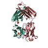 4f58C  3nh7S C: citing same article ( S: Starting model for refinement |
|---|---|
| Similar structure data |


-Links
 PDBj
PDBj

- Assembly
| Deposited unit | 
| ||||||||
|---|---|---|---|---|---|---|---|---|---|
| 1 |
| ||||||||
| Unit cell |
| ||||||||
| Components on special symmetry positions |
|
-Components
| #1: Antibody | Mass: 22482.826 Da / Num. of mol.: 1 / Fragment: light chain of L1 / Source method: isolated from a natural source / Source: (natural) Homo sapiens (human) |
|---|---|
| #2: Antibody | Mass: 24449.404 Da / Num. of mol.: 1 / Fragment: heavy chain of L1 / Source method: isolated from a natural source / Source: (natural) Homo sapiens (human) |
| #3: Chemical | ChemComp-GOL /   Mass: 92.094 Da / Num. of mol.: 1 / Source method: obtained synthetically / Formula: C3H8O3 Mass: 92.094 Da / Num. of mol.: 1 / Source method: obtained synthetically / Formula: C3H8O3 |
| #4: Water | ChemComp-HOH /  Mass: 18.015 Da / Num. of mol.: 532 / Source method: isolated from a natural source / Formula: H2O Mass: 18.015 Da / Num. of mol.: 532 / Source method: isolated from a natural source / Formula: H2O |
| Has protein modification | Y |
-Experimental details
-Experiment
| Experiment | Method: X-RAY DIFFRACTION / Number of used crystals: 1 |
|---|
- Sample preparation
| Crystal | Density Matthews: 2.58 Å3/Da / Density % sol: 52.27 % |
|---|---|
| Crystal grow | Temperature: 296 K / Method: vapor diffusion, hanging drop / pH: 6.5 Details: 0.17 M (NH4)2SO4, 0.085 M cacodylate pH 6.5, 25.5% (w/v) polyethylene glycol (PEG) 8000, and 15% (v/v) glycerol, VAPOR DIFFUSION, HANGING DROP, temperature 296K |
-Data collection
| Diffraction | Mean temperature: 100 K |
|---|---|
| Diffraction source | Source: SYNCHROTRON / Site: APS  / Beamline: 23-ID-D / Wavelength: 1.03319 Å / Beamline: 23-ID-D / Wavelength: 1.03319 Å |
| Detector | Type: MAR scanner 300 mm plate / Detector: IMAGE PLATE / Date: Nov 27, 2011 |
| Radiation | Monochromator: Mirrors / Protocol: SINGLE WAVELENGTH / Monochromatic (M) / Laue (L): M / Scattering type: x-ray |
| Radiation wavelength | Wavelength: 1.03319 Å / Relative weight: 1 |
| Reflection | Resolution: 1.7→41.5 Å / Num. all: 52788 / Num. obs: 50571 / % possible obs: 95.8 % / Observed criterion σ(F): 0 / Observed criterion σ(I): 0 |
| Reflection shell | Resolution: 1.7→1.73 Å / % possible all: 81.8 |
- Processing
| Software |
| |||||||||||||||||||||||||||||||||||||||||||||||||||||||||||||||||||||||||||||||||||||||||||||||||||||||||||||||||||||||||||||||||||||
|---|---|---|---|---|---|---|---|---|---|---|---|---|---|---|---|---|---|---|---|---|---|---|---|---|---|---|---|---|---|---|---|---|---|---|---|---|---|---|---|---|---|---|---|---|---|---|---|---|---|---|---|---|---|---|---|---|---|---|---|---|---|---|---|---|---|---|---|---|---|---|---|---|---|---|---|---|---|---|---|---|---|---|---|---|---|---|---|---|---|---|---|---|---|---|---|---|---|---|---|---|---|---|---|---|---|---|---|---|---|---|---|---|---|---|---|---|---|---|---|---|---|---|---|---|---|---|---|---|---|---|---|---|---|---|
| Refinement | Method to determine structure: MOLECULAR REPLACEMENT Starting model: 3NH7 Resolution: 1.7→41.5 Å / SU ML: 0.18 / σ(F): 1.36 / Phase error: 22.69 / Stereochemistry target values: ML
| |||||||||||||||||||||||||||||||||||||||||||||||||||||||||||||||||||||||||||||||||||||||||||||||||||||||||||||||||||||||||||||||||||||
| Solvent computation | Shrinkage radii: 0.6 Å / VDW probe radii: 0.9 Å / Solvent model: FLAT BULK SOLVENT MODEL / Bsol: 47.572 Å2 / ksol: 0.4 e/Å3 | |||||||||||||||||||||||||||||||||||||||||||||||||||||||||||||||||||||||||||||||||||||||||||||||||||||||||||||||||||||||||||||||||||||
| Displacement parameters |
| |||||||||||||||||||||||||||||||||||||||||||||||||||||||||||||||||||||||||||||||||||||||||||||||||||||||||||||||||||||||||||||||||||||
| Refinement step | Cycle: LAST / Resolution: 1.7→41.5 Å
| |||||||||||||||||||||||||||||||||||||||||||||||||||||||||||||||||||||||||||||||||||||||||||||||||||||||||||||||||||||||||||||||||||||
| Refine LS restraints |
| |||||||||||||||||||||||||||||||||||||||||||||||||||||||||||||||||||||||||||||||||||||||||||||||||||||||||||||||||||||||||||||||||||||
| LS refinement shell |
|
