 Movie
Movie Controller

[English] 日本語
 Yorodumi
- PDB-4emn: Crystal structure of RpfB catalytic domain in complex with benzamidine -
+ Open data

- Basic information
| Entry | Database: PDB / ID: 4emn | ||||||
|---|---|---|---|---|---|---|---|
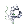
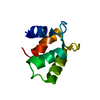
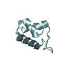
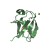
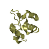
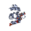
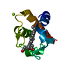
| Title | Crystal structure of RpfB catalytic domain in complex with benzamidine | ||||||
 Components Components | Probable resuscitation-promoting factor rpfB | ||||||
 Keywords Keywords | HYDROLASE / alpha beta | ||||||
| Function / homology |  Function and homology information Function and homology informationdormancy exit of symbiont in host / positive regulation of growth rate / Hydrolases / quorum sensing / regulation of cell population proliferation / hydrolase activity / negative regulation of gene expression / positive regulation of gene expression / extracellular region / plasma membrane Similarity search - Function | ||||||
| Biological species |   Mycobacterium tuberculosis (bacteria) Mycobacterium tuberculosis (bacteria) | ||||||
| Method |  X-RAY DIFFRACTION / SYNCHROTRON / MOLECULAR REPLACEMENT / Resolution: 1.17 Å X-RAY DIFFRACTION / SYNCHROTRON / MOLECULAR REPLACEMENT / Resolution: 1.17 Å | ||||||
 Authors Authors | Ruggiero, A. / Marchant, J. / Squeglia, F. / Makarov, V. / De Simone, A. / Berisio, R. | ||||||
 Citation Citation | Journal: J.Biomol.Struct.Dyn. / Year: 2013 Title: Molecular determinants of inactivation of the resuscitation promoting factor B from Mycobacterium tuberculosis. Authors: Ruggiero, A. / Marchant, J. / Squeglia, F. / Makarov, V. / De Simone, A. / Berisio, R. | ||||||
| History |
|
- Structure visualization
| Structure viewer | Molecule: MolmilJmol/JSmol |
|---|
- Downloads & links
-Download
| PDBx/mmCIF format | 4emn.cif.gz | 157.9 KB | Display | PDBx/mmCIF format |
|---|---|---|---|---|
| PDB format | pdb4emn.ent.gz | 125 KB | Display | PDB format |
| PDBx/mmJSON format | 4emn.json.gz | Tree view | PDBx/mmJSON format | |
| Others |  Other downloads Other downloads |
-Validation report
| Summary document | 4emn_validation.pdf.gz | 476.1 KB | Display | wwPDB validaton report |
|---|---|---|---|---|
| Full document | 4emn_full_validation.pdf.gz | 484.5 KB | Display | |
| Data in XML | 4emn_validation.xml.gz | 22.8 KB | Display | |
| Data in CIF | 4emn_validation.cif.gz | 33.4 KB | Display | |
| Arichive directory | https://data.pdbj.org/pub/pdb/validation_reports/em/4emnftp://data.pdbj.org/pub/pdb/validation_reports/em/4emn | HTTPS FTP |
-Related structure data
| Related structure data |  3eo5S S: Starting model for refinement |
|---|---|
| Similar structure data |


-Links
 PDBj
PDBj


- Assembly
| Deposited unit | 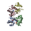
| ||||||||
|---|---|---|---|---|---|---|---|---|---|
| 1 | 
| ||||||||
| 2 | 
| ||||||||
| 3 | 
| ||||||||
| 4 | 
| ||||||||
| Unit cell |
|
-Components
| #1: Protein | Mass: 8591.399 Da / Num. of mol.: 4 / Fragment: catalytic domain Source method: isolated from a genetically manipulated source Source: (gene. exp.) Mycobacterium tuberculosis (bacteria) / Gene: MT1038, rpfB, Rv1009 / Production host: #2: Chemical | ChemComp-BEN /   Mass: 120.152 Da / Num. of mol.: 6 / Source method: obtained synthetically / Formula: C7H8N2 Mass: 120.152 Da / Num. of mol.: 6 / Source method: obtained synthetically / Formula: C7H8N2#3: Chemical |   Mass: 96.063 Da / Num. of mol.: 2 / Source method: obtained synthetically / Formula: SO4 Mass: 96.063 Da / Num. of mol.: 2 / Source method: obtained synthetically / Formula: SO4#4: Water | ChemComp-HOH / |  Mass: 18.015 Da / Num. of mol.: 567 / Source method: isolated from a natural source / Formula: H2O Mass: 18.015 Da / Num. of mol.: 567 / Source method: isolated from a natural source / Formula: H2OHas protein modification | Y | |
|---|
-Experimental details
-Experiment
| Experiment | Method: X-RAY DIFFRACTION / Number of used crystals: 1 |
|---|
- Sample preparation
| Crystal | Density Matthews: 2.02 Å3/Da / Density % sol: 39.2 % |
|---|---|
| Crystal grow | Temperature: 293 K / Method: vapor diffusion / pH: 7.5 Details: 0.5% peg8000, 1M ammonium sulfate, pH 7.5, VAPOR DIFFUSION, temperature 293K |
-Data collection
| Diffraction | Mean temperature: 100 K |
|---|---|
| Diffraction source | Source: SYNCHROTRON / Site: ELETTRA  / Beamline: 5.2R / Wavelength: 1 Å / Beamline: 5.2R / Wavelength: 1 Å |
| Detector | Type: MAR CCD 165 mm / Detector: CCD / Date: Jan 1, 2008 |
| Radiation | Monochromator: GRAPHITE / Protocol: SINGLE WAVELENGTH / Monochromatic (M) / Laue (L): M / Scattering type: x-ray |
| Radiation wavelength | Wavelength: 1 Å / Relative weight: 1 |
| Reflection | Resolution: 1.17→30 Å / Num. obs: 85254 / % possible obs: 92 % / Observed criterion σ(F): 0 / Observed criterion σ(I): 0 / Redundancy: 3.1 % / Rmerge(I) obs: 0.052 |
| Reflection shell | Resolution: 1.17→1.21 Å / Rmerge(I) obs: 0.021 / % possible all: 75.1 |
- Processing
| Software |
| ||||||||||||||||||||
|---|---|---|---|---|---|---|---|---|---|---|---|---|---|---|---|---|---|---|---|---|---|
| Refinement | Method to determine structure: MOLECULAR REPLACEMENT Starting model: PDB entry 3eo5 Resolution: 1.17→15 Å / σ(F): 4 / Stereochemistry target values: Engh & Huber
| ||||||||||||||||||||
| Refine analyze | Luzzati coordinate error obs: 0.1 Å | ||||||||||||||||||||
| Refinement step | Cycle: LAST / Resolution: 1.17→15 Å
| ||||||||||||||||||||
| Refine LS restraints |
| ||||||||||||||||||||
| LS refinement shell | Resolution: 1.17→1.21 Å
|
