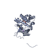
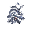
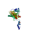
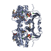
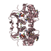
- PDB-4ej7: Crystal structure of the aminoglycoside phosphotransferase APH(3'... -
+
Open data
ID or keywords:
Loading...
-
Basic information
Entry
Database: PDB / ID: 4ej7
Title
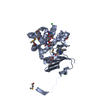
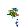
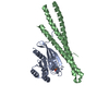
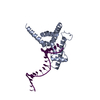
Crystal structure of the aminoglycoside phosphotransferase APH(3')-Ia, ATP-bound
Components
Aminoglycoside 3'-phosphotransferase AphA1-IAB
Keywords
TRANSFERASE / CENTER FOR STRUCTURAL GENOMICS OF INFECTIOUS DISEASES / CSGID / NIAID / National Institute of Allergy and Infectious Diseases / EUKARYOTIC PROTEIN KINASE-LIKE FOLD / ALPHA/BETA PROTEIN / PHOSPHOTRANSFERASE / AMINOGLYCOSIDE PHOSPHOTRANSFERASE / ANTIBIOTIC RESISTANCE / AMINOGLYCOSIDES / KANAMYCIN / GTP / INTRACELLULAR
Function / homology
Function and homology information
kanamycin kinase / kanamycin kinase activity / phosphorylation / response to antibiotic / ATP binding / metal ion binding Similarity search - Function
Mass: 18.015 Da / Num. of mol.: 294 / Source method: isolated from a natural source / Formula: H2O
Has protein modification
Y
-
Experimental details
-
Experiment
Experiment
Method: X-RAY DIFFRACTION / Number of used crystals: 1
-
Sample preparation
Crystal
Density Matthews: 2.66 Å3/Da / Density % sol: 53.73 %
Crystal grow
Temperature: 298 K / Method: vapor diffusion, sitting drop / pH: 7 Details: 0.1 M CA ACETATE, 16% PEG3350, 2 MM ATP, pH 7, VAPOR DIFFUSION, SITTING DROP, temperature 298K
Monochromator: GRAPHITE / Protocol: SINGLE WAVELENGTH / Monochromatic (M) / Laue (L): M / Scattering type: x-ray
Radiation wavelength
Wavelength: 0.9794 Å / Relative weight: 1
Reflection
Resolution: 2.29→43.669 Å / Num. obs: 91198 / % possible obs: 91.9 % / Observed criterion σ(F): 0 / Observed criterion σ(I): 1 / Net I/σ(I): 12.552
Reflection shell
Resolution: 2.29→2.3 Å / Mean I/σ(I) obs: 1.5 / Rsym value: 0.5298 / % possible all: 69.4
-
Processing
Software
Name
Version
Classification
ADSC
Quantum
datacollection
SHELXS
phasing
PHENIX
(phenix.refine: 1.7.3_928)
refinement
HKL-2000
datareduction
HKL-2000
datascaling
Refinement
Method to determine structure: SAD / Resolution: 2.29→43.669 Å / SU ML: 0.38 / Cross valid method: THROUGHOUT / σ(F): 1.34 / Phase error: 29.86 / Stereochemistry target values: ML
Rfactor
Num. reflection
% reflection
Selection details
Rfree
0.2516
3790
4.19 %
random
Rwork
0.2134
-
-
-
obs
0.215
90409
96.27 %
-
Solvent computation
Shrinkage radii: 0.9 Å / VDW probe radii: 1.11 Å / Solvent model: FLAT BULK SOLVENT MODEL / Bsol: 29.351 Å2 / ksol: 0.321 e/Å3
Displacement parameters
Baniso -1
Baniso -2
Baniso -3
1-
4.8067 Å2
-0 Å2
0 Å2
2-
-
31.4101 Å2
-0 Å2
3-
-
-
20.1465 Å2
Refinement step
Cycle: LAST / Resolution: 2.29→43.669 Å
Protein
Nucleic acid
Ligand
Solvent
Total
Num. atoms
6472
0
112
294
6878
Refine LS restraints
Refine-ID
Type
Dev ideal
Number
X-RAY DIFFRACTION
f_bond_d
0.009
6879
X-RAY DIFFRACTION
f_angle_d
0.694
9395
X-RAY DIFFRACTION
f_dihedral_angle_d
12.75
2535
X-RAY DIFFRACTION
f_chiral_restr
0.043
980
X-RAY DIFFRACTION
f_plane_restr
0.003
1215
LS refinement shell
Resolution (Å)
Rfactor Rfree
Num. reflection Rfree
Rfactor Rwork
Num. reflection Rwork
Refine-ID
% reflection obs (%)
2.2903-2.3722
0.3448
359
0.3205
8391
X-RAY DIFFRACTION
93
2.3722-2.4672
0.3291
383
0.2955
8699
X-RAY DIFFRACTION
97
2.4672-2.5794
0.3729
381
0.2927
8801
X-RAY DIFFRACTION
97
2.5794-2.7154
0.3343
382
0.2662
8735
X-RAY DIFFRACTION
97
2.7154-2.8855
0.3103
388
0.2519
8662
X-RAY DIFFRACTION
97
2.8855-3.1082
0.2985
383
0.2502
8756
X-RAY DIFFRACTION
97
3.1082-3.4209
0.3134
391
0.2232
8656
X-RAY DIFFRACTION
97
3.4209-3.9157
0.2244
375
0.201
8768
X-RAY DIFFRACTION
97
3.9157-4.9323
0.2131
379
0.1634
8650
X-RAY DIFFRACTION
96
4.9323-43.6765
0.1933
369
0.1961
8501
X-RAY DIFFRACTION
94
Refinement TLS params.
Method: refined / Refine-ID: X-RAY DIFFRACTION
ID
L11 (°2)
L12 (°2)
L13 (°2)
L22 (°2)
L23 (°2)
L33 (°2)
S11 (Å °)
S12 (Å °)
S13 (Å °)
S21 (Å °)
S22 (Å °)
S23 (Å °)
S31 (Å °)
S32 (Å °)
S33 (Å °)
T11 (Å2)
T12 (Å2)
T13 (Å2)
T22 (Å2)
T23 (Å2)
T33 (Å2)
Origin x (Å)
Origin y (Å)
Origin z (Å)
1
0.1426
0.0236
0.0704
0.0644
-0.0114
0.1188
0.093
0.0342
-0.4693
-0.186
0.1506
-0.034
-0.065
-0.0895
0.2115
-0.1448
0.187
-0.24
0.0749
0.156
0.1742
84.1093
25.0009
47.5867
2
0.1336
-0.11
0.0046
0.1338
0.0415
0.1559
-0.028
-0.08
-0.0447
-0.0171
0.0761
-0.03
-0.0155
-0.149
0.0039
0.2077
-0.0066
0.0229
0.1811
-0.0016
0.1512
73.9944
47.6784
56.6634
3
0.1989
-0.0059
0.167
0.1977
-0.0443
0.4835
0.3711
0.1106
-0.18
-0.15
0.2106
0.3177
0.1586
-0.0572
0.3098
0.2063
0.0024
-0.0866
0.2145
0.1775
0.3849
33.1321
73.401
75.7129
4
0.0862
-0.1129
-0.0835
0.0738
0.082
0.0971
0.0668
0.0498
-0.0066
0.0435
-0.0737
0.1161
0.1217
0.0347
-0.0006
0.2774
0.0263
-0.0133
0.1835
-0.0127
0.1722
56.0076
64.4034
67.8521
5
0.0509
-0.0249
0.0156
0.0803
-0.026
0.1734
0.1806
0.0408
0.1764
-0.0386
-0.0686
0.0003
0.1725
-0.0459
0.0152
0.1674
0.0039
0.0063
0.2642
-0.017
0.2219
44.0698
29.7781
47.9276
6
0.1241
0.0377
0.0483
0.1216
0.0324
0.1298
0.1276
-0.2442
-0.4124
0.0038
-0.0391
-0.0535
0.0094
0.0997
0.0834
0.2703
-0.0692
-0.1513
0.3143
0.0673
0.2764
54.1111
7.0148
56.7389
Refinement TLS group
ID
Refine-ID
Refine TLS-ID
Selection details
1
X-RAY DIFFRACTION
1
chainAandresid1:102
2
X-RAY DIFFRACTION
2
chainAandresid103:271
3
X-RAY DIFFRACTION
3
chainBandresid1:102
4
X-RAY DIFFRACTION
4
chainBandresid103:271
5
X-RAY DIFFRACTION
5
chainCandresid1:102
6
X-RAY DIFFRACTION
6
chainCandresid103:271
+
About Yorodumi
-
News
-
Feb 9, 2022. New format data for meta-information of EMDB entries
New format data for meta-information of EMDB entries
Version 3 of the EMDB header file is now the official format.
The previous official version 1.9 will be removed from the archive.
In the structure databanks used in Yorodumi, some data are registered as the other names, "COVID-19 virus" and "2019-nCoV". Here are the details of the virus and the list of structure data.
Jan 31, 2019. EMDB accession codes are about to change! (news from PDBe EMDB page)
EMDB accession codes are about to change! (news from PDBe EMDB page)
The allocation of 4 digits for EMDB accession codes will soon come to an end. Whilst these codes will remain in use, new EMDB accession codes will include an additional digit and will expand incrementally as the available range of codes is exhausted. The current 4-digit format prefixed with “EMD-” (i.e. EMD-XXXX) will advance to a 5-digit format (i.e. EMD-XXXXX), and so on. It is currently estimated that the 4-digit codes will be depleted around Spring 2019, at which point the 5-digit format will come into force.
The EM Navigator/Yorodumi systems omit the EMD- prefix.
Related info.:Q: What is EMD? / ID/Accession-code notation in Yorodumi/EM Navigator
Yorodumi is a browser for structure data from EMDB, PDB, SASBDB, etc.
This page is also the successor to EM Navigator detail page, and also detail information page/front-end page for Omokage search.
The word "yorodu" (or yorozu) is an old Japanese word meaning "ten thousand". "mi" (miru) is to see.
Related info.:EMDB / PDB / SASBDB / Comparison of 3 databanks / Yorodumi Search / Aug 31, 2016. New EM Navigator & Yorodumi / Yorodumi Papers / Jmol/JSmol / Function and homology information / Changes in new EM Navigator and Yorodumi
 Movie
Movie Controller

 Yorodumi
 Open data
 Basic information
 Components
 Keywords
 Function and homology information
 Acinetobacter baumannii (bacteria)
 X-RAY DIFFRACTION /
  Authors
 Citation
 Structure visualization
 Downloads & links
 Other downloads
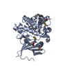


 PDBj
PDBj
 Assembly


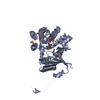
 Mass: 507.181 Da / Num. of mol.: 3 / Source method: obtained synthetically / Formula: C10H16N5O13P3 / Comment: ATP, energy-carrying molecule*YM
 Mass: 40.078 Da / Num. of mol.: 12 / Source method: obtained synthetically / Formula: Ca
 Mass: 106.120 Da / Num. of mol.: 1 / Source method: obtained synthetically / Formula: C4H10O3
 Mass: 18.015 Da / Num. of mol.: 294 / Source method: isolated from a natural source / Formula: H2O
 Sample preparation
 / Beamline: 19-ID / Wavelength: 0.9794 Å
 Processing