 Movie
Movie Controller

[English] 日本語
 Yorodumi
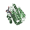
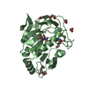
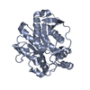
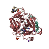
- PDB-4e46: Structure of Rhodococcus rhodochrous haloalkane dehalogenase DhaA... -
+ Open data

- Basic information
| Entry | Database: PDB / ID: 4.0E+46 | ||||||
|---|---|---|---|---|---|---|---|
| Title | Structure of Rhodococcus rhodochrous haloalkane dehalogenase DhaA in complex with 2-propanol | ||||||
 Components Components | Haloalkane dehalogenase | ||||||
 Keywords Keywords | HYDROLASE / catalytic pentad / alpha/beta hydrolase fold / halide binding / hydrolytic dehalogenation | ||||||
| Function / homology |  Function and homology information Function and homology informationhaloalkane dehalogenase / haloalkane dehalogenase activity / response to toxic substance / membrane Similarity search - Function | ||||||
| Biological species |  Rhodococcus rhodochrous (bacteria) Rhodococcus rhodochrous (bacteria) | ||||||
| Method |  X-RAY DIFFRACTION / SYNCHROTRON / MOLECULAR REPLACEMENT / Resolution: 1.26 Å X-RAY DIFFRACTION / SYNCHROTRON / MOLECULAR REPLACEMENT / Resolution: 1.26 Å | ||||||
 Authors Authors | Stsiapanava, A. / Chaloupkova, R. / Damborsky, J. / Kuta Smatanova, I. | ||||||
 Citation Citation | Journal: Chembiochem / Year: 2013 Title: Expansion of access tunnels and active-site cavities influence activity of haloalkane dehalogenases in organic cosolvents. Authors: Stepankova, V. / Khabiri, M. / Brezovsky, J. / Pavelka, A. / Sykora, J. / Amaro, M. / Minofar, B. / Prokop, Z. / Hof, M. / Ettrich, R. / Chaloupkova, R. / Damborsky, J. | ||||||
| History |
|
- Structure visualization
| Structure viewer | Molecule: MolmilJmol/JSmol |
|---|
- Downloads & links
-Download
| PDBx/mmCIF format | 4e46.cif.gz | 147.4 KB | Display | PDBx/mmCIF format |
|---|---|---|---|---|
| PDB format | pdb4e46.ent.gz | 112.4 KB | Display | PDB format |
| PDBx/mmJSON format | 4e46.json.gz | Tree view | PDBx/mmJSON format | |
| Others |  Other downloads Other downloads |
-Validation report
| Arichive directory | https://data.pdbj.org/pub/pdb/validation_reports/e4/4e46ftp://data.pdbj.org/pub/pdb/validation_reports/e4/4e46 | HTTPS FTP |
|---|
-Related structure data
| Related structure data |  3fbwS S: Starting model for refinement |
|---|---|
| Similar structure data |


-Links
 PDBj
PDBj


- Assembly
| Deposited unit | 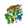
| ||||||||
|---|---|---|---|---|---|---|---|---|---|
| 1 |
| ||||||||
| Unit cell |
|
-Components
| #1: Protein | Mass: 33696.434 Da / Num. of mol.: 1 Source method: isolated from a genetically manipulated source Source: (gene. exp.) Rhodococcus rhodochrous (bacteria) / Strain: NCIB 13064 / Gene: dhaA / Plasmid: pAQN / Production host: | ||||
|---|---|---|---|---|---|
| #2: Chemical | ChemComp-IPA /   Mass: 60.095 Da / Num. of mol.: 1 / Source method: obtained synthetically / Formula: C3H8O Mass: 60.095 Da / Num. of mol.: 1 / Source method: obtained synthetically / Formula: C3H8O | ||||
| #3: Chemical |   Mass: 59.044 Da / Num. of mol.: 2 / Source method: obtained synthetically / Formula: C2H3O2 Mass: 59.044 Da / Num. of mol.: 2 / Source method: obtained synthetically / Formula: C2H3O2#4: Chemical | ChemComp-CL / |   Mass: 35.453 Da / Num. of mol.: 1 / Source method: obtained synthetically / Formula: Cl Mass: 35.453 Da / Num. of mol.: 1 / Source method: obtained synthetically / Formula: Cl#5: Water | ChemComp-HOH / |  Mass: 18.015 Da / Num. of mol.: 542 / Source method: isolated from a natural source / Formula: H2O Mass: 18.015 Da / Num. of mol.: 542 / Source method: isolated from a natural source / Formula: H2O |
-Experimental details
-Experiment
| Experiment | Method: X-RAY DIFFRACTION / Number of used crystals: 1 |
|---|
- Sample preparation
| Crystal | Density Matthews: 2.1 Å3/Da / Density % sol: 41.34 % |
|---|---|
| Crystal grow | Temperature: 277 K / Method: vapor diffusion, sitting drop Details: 100 mM sodium acetate, 24% PEG4000, 11% 2-propanol, VAPOR DIFFUSION, SITTING DROP, temperature 277K |
-Data collection
| Diffraction | Mean temperature: 100 K |
|---|---|
| Diffraction source | Source: SYNCHROTRON / Site: EMBL/DESY, HAMBURG  / Beamline: X12 / Wavelength: 1.033 Å / Beamline: X12 / Wavelength: 1.033 Å |
| Detector | Type: MARMOSAIC 225 mm CCD / Detector: CCD / Date: Mar 25, 2009 |
| Radiation | Monochromator: double crystal Si(111), horizontally focusing Protocol: SINGLE WAVELENGTH / Monochromatic (M) / Laue (L): M / Scattering type: x-ray |
| Radiation wavelength | Wavelength: 1.033 Å / Relative weight: 1 |
| Reflection | Resolution: 1.26→50 Å / Num. all: 69524 / Num. obs: 65492 / % possible obs: 94.3 % / Observed criterion σ(F): 0 / Observed criterion σ(I): 0 / Redundancy: 2.4 % / Biso Wilson estimate: 13.3 Å2 / Rmerge(I) obs: 0.037 / Net I/σ(I): 29.9 |
| Reflection shell | Resolution: 1.26→1.31 Å / Redundancy: 2.1 % / Rmerge(I) obs: 0.113 / Mean I/σ(I) obs: 6.8 / % possible all: 90.2 |
- Processing
| Software |
| |||||||||||||||||||||||||||||||||
|---|---|---|---|---|---|---|---|---|---|---|---|---|---|---|---|---|---|---|---|---|---|---|---|---|---|---|---|---|---|---|---|---|---|---|
| Refinement | Method to determine structure: MOLECULAR REPLACEMENT Starting model: PDB ENTRY 3FBW Resolution: 1.26→10 Å / Num. parameters: 24815 / Num. restraintsaints: 30840 Isotropic thermal model: MIXED ISOTROPIC AND ANISOTROPIC THERMAL MODEL σ(F): 0 / Stereochemistry target values: ENGH AND HUBER Details: STRUCTURE SOLUTION AND REFINEMENT WERE PERFORMED USING FREE R FOR CROSS-VALIDATION THROUGHOUT: FREE R = 0.150 FROM A RANDOM TEST SET COMPRISING 5% OF REFLECTIONS (3476 TOTAL). FINAL ...Details: STRUCTURE SOLUTION AND REFINEMENT WERE PERFORMED USING FREE R FOR CROSS-VALIDATION THROUGHOUT: FREE R = 0.150 FROM A RANDOM TEST SET COMPRISING 5% OF REFLECTIONS (3476 TOTAL). FINAL REFINEMENT WAS PERFORMED USING ALL REFLECTIONS.
| |||||||||||||||||||||||||||||||||
| Displacement parameters | Biso mean: 16 Å2 | |||||||||||||||||||||||||||||||||
| Refine analyze | Num. disordered residues: 49 / Occupancy sum hydrogen: 2134 / Occupancy sum non hydrogen: 2902.45 | |||||||||||||||||||||||||||||||||
| Refinement step | Cycle: LAST / Resolution: 1.26→10 Å
| |||||||||||||||||||||||||||||||||
| Refine LS restraints |
|
