 Movie
Movie Controller

[English] 日本語
 Yorodumi
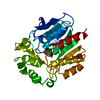
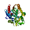
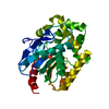
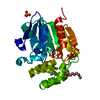
- PDB-1cqw: NAI COCRYSTALLISED WITH HALOALKANE DEHALOGENASE FROM A RHODOCOCCU... -
+ Open data

- Basic information
| Entry | Database: PDB / ID: 1cqw | ||||||
|---|---|---|---|---|---|---|---|
| Title | NAI COCRYSTALLISED WITH HALOALKANE DEHALOGENASE FROM A RHODOCOCCUS SPECIES | ||||||
 Components Components | HALOALKANE DEHALOGENASE; 1-CHLOROHEXANE HALIDOHYDROLASE | ||||||
 Keywords Keywords | HYDROLASE / A/B HYDROLASE FOLD / DEHALOGENASE I-S BOND | ||||||
| Function / homology |  Function and homology information Function and homology information | ||||||
| Biological species |  Rhodococcus sp. (bacteria) Rhodococcus sp. (bacteria) | ||||||
| Method |  X-RAY DIFFRACTION / SYNCHROTRON / Resolution: 1.5 Å X-RAY DIFFRACTION / SYNCHROTRON / Resolution: 1.5 Å | ||||||
 Authors Authors | Newman, J. / Peat, T.S. / Richard, R. / Kan, L. / Swanson, P.E. / Affholter, J.A. / Holmes, I.H. / Schindler, J.F. / Unkefer, C.J. / Terwilliger, T.C. | ||||||
 Citation Citation | Journal: Biochemistry / Year: 1999 Title: Haloalkane dehalogenases: structure of a Rhodococcus enzyme. Authors: Newman, J. / Peat, T.S. / Richard, R. / Kan, L. / Swanson, P.E. / Affholter, J.A. / Holmes, I.H. / Schindler, J.F. / Unkefer, C.J. / Terwilliger, T.C. | ||||||
| History |
|
- Structure visualization
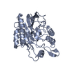
| Structure viewer | Molecule: MolmilJmol/JSmol |
|---|
- Downloads & links
-Download
| PDBx/mmCIF format | 1cqw.cif.gz | 80.7 KB | Display | PDBx/mmCIF format |
|---|---|---|---|---|
| PDB format | pdb1cqw.ent.gz | 60.5 KB | Display | PDB format |
| PDBx/mmJSON format | 1cqw.json.gz | Tree view | PDBx/mmJSON format | |
| Others |  Other downloads Other downloads |
-Validation report
| Arichive directory | https://data.pdbj.org/pub/pdb/validation_reports/cq/1cqwftp://data.pdbj.org/pub/pdb/validation_reports/cq/1cqw | HTTPS FTP |
|---|
-Related structure data


-Links
 PDBj
PDBj


- Assembly
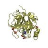
| Deposited unit | 
| ||||||||
|---|---|---|---|---|---|---|---|---|---|
| 1 |
| ||||||||
| Unit cell |
|
-Components
| #1: Protein | Mass: 33334.059 Da / Num. of mol.: 1 Source method: isolated from a genetically manipulated source Details: COCRYSTALLIZED WITH NAI / Source: (gene. exp.) Rhodococcus sp. (bacteria) / Plasmid: PTRCHIS / Production host: | ||
|---|---|---|---|
| #2: Chemical |   Mass: 126.904 Da / Num. of mol.: 2 / Source method: obtained synthetically / Formula: I Mass: 126.904 Da / Num. of mol.: 2 / Source method: obtained synthetically / Formula: I#3: Water | ChemComp-HOH / |  Mass: 18.015 Da / Num. of mol.: 394 / Source method: isolated from a natural source / Formula: H2O Mass: 18.015 Da / Num. of mol.: 394 / Source method: isolated from a natural source / Formula: H2O |
-Experimental details
-Experiment
| Experiment | Method: X-RAY DIFFRACTION / Number of used crystals: 1 |
|---|
- Sample preparation
| Crystal | Density Matthews: 2.34 Å3/Da / Density % sol: 47.51 % | ||||||||||||||||||||||||||||||||||||||||
|---|---|---|---|---|---|---|---|---|---|---|---|---|---|---|---|---|---|---|---|---|---|---|---|---|---|---|---|---|---|---|---|---|---|---|---|---|---|---|---|---|---|
| Crystal grow | Temperature: 281 K / Method: vapor diffusion, hanging drop / pH: 5.5 Details: 20-25% PEG 1.5K, 0.1M MES pH 5.5 0.3M NaAcetate, VAPOR DIFFUSION, HANGING DROP, temperature 281K | ||||||||||||||||||||||||||||||||||||||||
| Crystal grow | *PLUS Temperature: 8 ℃ / pH: 7.5 | ||||||||||||||||||||||||||||||||||||||||
| Components of the solutions | *PLUS
|
-Data collection
| Diffraction | Mean temperature: 110 K |
|---|---|
| Diffraction source | Source: SYNCHROTRON / Site: NSLS  / Beamline: X8C / Wavelength: 1.072 / Beamline: X8C / Wavelength: 1.072 |
| Detector | Type: MARRESEARCH / Detector: AREA DETECTOR / Date: Nov 1, 1997 |
| Radiation | Protocol: SINGLE WAVELENGTH / Monochromatic (M) / Laue (L): M / Scattering type: x-ray |
| Radiation wavelength | Wavelength: 1.072 Å / Relative weight: 1 |
| Reflection | Resolution: 1.5→12.9 Å / Num. all: 222118 / Num. obs: 47965 / % possible obs: 93.1 % / Observed criterion σ(F): 0 / Observed criterion σ(I): 3 / Redundancy: 4.2 % / Biso Wilson estimate: 13.2 Å2 / Rmerge(I) obs: 0.065 / Net I/σ(I): 10.6 |
| Reflection shell | Resolution: 1.49→1.57 Å / Redundancy: 3.2 % / Rmerge(I) obs: 0.154 / Num. unique all: 5826 / % possible all: 78.6 |
| Reflection | *PLUS Num. measured all: 222118 / Rmerge(I) obs: 0.046 |
| Reflection shell | *PLUS % possible obs: 75.6 % |
- Processing
| Software |
| ||||||||||||||||||||||||||||||||||||
|---|---|---|---|---|---|---|---|---|---|---|---|---|---|---|---|---|---|---|---|---|---|---|---|---|---|---|---|---|---|---|---|---|---|---|---|---|---|
| Refinement | Resolution: 1.5→12.9 Å / Rfactor Rfree error: 0.004 / Data cutoff high absF: 1371216.83 / Data cutoff low absF: 0 / Isotropic thermal model: RESTRAINED / Cross valid method: THROUGHOUT / σ(F): 3
| ||||||||||||||||||||||||||||||||||||
| Displacement parameters | Biso mean: 12.1 Å2
| ||||||||||||||||||||||||||||||||||||
| Refine analyze |
| ||||||||||||||||||||||||||||||||||||
| Refinement step | Cycle: LAST / Resolution: 1.5→12.9 Å
| ||||||||||||||||||||||||||||||||||||
| Refine LS restraints |
| ||||||||||||||||||||||||||||||||||||
| LS refinement shell | Resolution: 1.5→1.59 Å / Rfactor Rfree error: 0.011 / Total num. of bins used: 6
| ||||||||||||||||||||||||||||||||||||
| Xplor file |
| ||||||||||||||||||||||||||||||||||||
| Software | *PLUS Name: CNS / Version: 0.3 / Classification: refinement | ||||||||||||||||||||||||||||||||||||
| Refinement | *PLUS Rfactor obs: 0.175 / Rfactor Rfree: 0.187 / Rfactor Rwork: 0.175 | ||||||||||||||||||||||||||||||||||||
| Solvent computation | *PLUS | ||||||||||||||||||||||||||||||||||||
| Displacement parameters | *PLUS | ||||||||||||||||||||||||||||||||||||
| Refine LS restraints | *PLUS
|
