 Movie
Movie Controller

[English] 日本語
 Yorodumi
- PDB-3i5v: Crystal structure of beta toxin 275-280 from Staphylococcus aureus -
+ Open data

- Basic information
| Entry | Database: PDB / ID: 3i5v | ||||||
|---|---|---|---|---|---|---|---|
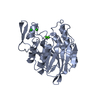
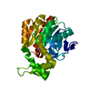
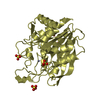
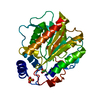
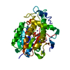
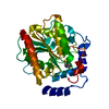
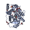
| Title | Crystal structure of beta toxin 275-280 from Staphylococcus aureus | ||||||
 Components Components | Beta-hemolysin | ||||||
 Keywords Keywords | TOXIN / beta toxin / hemolysin / sphingomyelinase | ||||||
| Function / homology |  Function and homology information Function and homology informationsphingomyelin phosphodiesterase activity / phospholipase C / phosphatidylcholine phospholipase C activity / toxin activity / killing of cells of another organism / extracellular region / metal ion binding Similarity search - Function | ||||||
| Biological species |   Staphylococcus aureus (bacteria) Staphylococcus aureus (bacteria) | ||||||
| Method |  X-RAY DIFFRACTION / SYNCHROTRON / MOLECULAR REPLACEMENT / Resolution: 2.8 Å X-RAY DIFFRACTION / SYNCHROTRON / MOLECULAR REPLACEMENT / Resolution: 2.8 Å | ||||||
 Authors Authors | Huseby, M. / Shi, K. / Kruse, A.C. / Ohlendorf, D.H. | ||||||
 Citation Citation | Journal: To be Published Title: Structure and biological functions of beta toxin from Staphylococcus aureus: Role of the hydrophobic beta hairpin in virulence Authors: Huseby, M. / Shi, K. / Kruse, A.C. / Digre, J. / Mengistu, F. / Bohach, G.A. / Schlievert, P.S. / Ohlendorf, D.H. / Earhart, C.A. #1: Journal: J.Bacteriol. / Year: 2007 Title: Structure and biological activities of beta toxin from Staphylococcus aureus. Authors: Huseby, M. / Shi, K. / Brown, C.K. / Digre, J. / Mengistu, F. / Seo, K.S. / Bohach, G.A. / Schlievert, P.M. / Ohlendorf, D.H. / Earhart, C.A. | ||||||
| History |
|
- Structure visualization
| Structure viewer | Molecule: MolmilJmol/JSmol |
|---|
- Downloads & links
-Download
| PDBx/mmCIF format | 3i5v.cif.gz | 239.2 KB | Display | PDBx/mmCIF format |
|---|---|---|---|---|
| PDB format | pdb3i5v.ent.gz | 191.5 KB | Display | PDB format |
| PDBx/mmJSON format | 3i5v.json.gz | Tree view | PDBx/mmJSON format | |
| Others |  Other downloads Other downloads |
-Validation report
| Arichive directory | https://data.pdbj.org/pub/pdb/validation_reports/i5/3i5vftp://data.pdbj.org/pub/pdb/validation_reports/i5/3i5v | HTTPS FTP |
|---|
-Related structure data


-Links
 PDBj
PDBj


- Assembly
| Deposited unit | 
| ||||||||
|---|---|---|---|---|---|---|---|---|---|
| 1 | 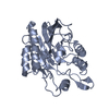
| ||||||||
| 2 | 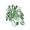
| ||||||||
| 3 | 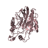
| ||||||||
| 4 | 
| ||||||||
| Unit cell |
|
-Components
| #1: Protein | Mass: 35561.543 Da / Num. of mol.: 4 / Fragment: UNP residues 35-330 Source method: isolated from a genetically manipulated source Source: (gene. exp.) Staphylococcus aureus (bacteria) / Strain: RN4220 / Gene: hlb / Plasmid: pET28b / Production host: #2: Chemical | ChemComp-DGA / |   Mass: 625.018 Da / Num. of mol.: 1 / Source method: obtained synthetically / Formula: C39H76O5 Mass: 625.018 Da / Num. of mol.: 1 / Source method: obtained synthetically / Formula: C39H76O5#3: Water | ChemComp-HOH / |  Mass: 18.015 Da / Num. of mol.: 116 / Source method: isolated from a natural source / Formula: H2O Mass: 18.015 Da / Num. of mol.: 116 / Source method: isolated from a natural source / Formula: H2OHas protein modification | Y | Sequence details | THIS IS A DELETION MUTANT DELTA(308-313)/DG, IN WHICH RESIDUES 308-313 HAVE BEEN DELETED AND ...THIS IS A DELETION MUTANT DELTA(308-313)/DG, IN WHICH RESIDUES 308-313 HAVE BEEN DELETED AND REPLACED WITH RESIDUES ASP-GLY. | |
|---|
-Experimental details
-Experiment
| Experiment | Method: X-RAY DIFFRACTION / Number of used crystals: 1 |
|---|
- Sample preparation
| Crystal | Density Matthews: 2.02 Å3/Da / Density % sol: 38.97 % |
|---|---|
| Crystal grow | Temperature: 291 K / Method: vapor diffusion, sitting drop / pH: 7 Details: 0.15-0.25 M NaF, 28-34% PEG 3350, pH 7.0, VAPOR DIFFUSION, SITTING DROP, temperature 291K |
-Data collection
| Diffraction | Mean temperature: 100 K |
|---|---|
| Diffraction source | Source: SYNCHROTRON / Site: APS  / Beamline: 14-BM-C / Wavelength: 0.9002 Å / Beamline: 14-BM-C / Wavelength: 0.9002 Å |
| Detector | Type: ADSC QUANTUM 315 / Detector: CCD / Date: Feb 10, 2008 / Details: mirrors |
| Radiation | Monochromator: bent Ge(111) / Protocol: SINGLE WAVELENGTH / Monochromatic (M) / Laue (L): M / Scattering type: x-ray |
| Radiation wavelength | Wavelength: 0.9002 Å / Relative weight: 1 |
| Reflection | Resolution: 2.8→38.4 Å / Num. all: 22252 / Num. obs: 22252 / % possible obs: 94.97 % / Observed criterion σ(I): 0 / Biso Wilson estimate: 38.626 Å2 |
| Reflection shell | Resolution: 2.8→2.99 Å / % possible all: 38.8 |
- Processing
| Software |
| |||||||||||||||||||||||||||||||||||||||||||||||||||||||||||||||||||||||||||||||||||||||||||||||||||||||||||||||||||||||||||||
|---|---|---|---|---|---|---|---|---|---|---|---|---|---|---|---|---|---|---|---|---|---|---|---|---|---|---|---|---|---|---|---|---|---|---|---|---|---|---|---|---|---|---|---|---|---|---|---|---|---|---|---|---|---|---|---|---|---|---|---|---|---|---|---|---|---|---|---|---|---|---|---|---|---|---|---|---|---|---|---|---|---|---|---|---|---|---|---|---|---|---|---|---|---|---|---|---|---|---|---|---|---|---|---|---|---|---|---|---|---|---|---|---|---|---|---|---|---|---|---|---|---|---|---|---|---|---|
| Refinement | Method to determine structure: MOLECULAR REPLACEMENT Starting model: Wild-type beta toxin from Staphylococcus aureus Resolution: 2.8→38.4 Å / Occupancy max: 1 / Occupancy min: 1 / Isotropic thermal model: Isotropic / σ(F): 0 / σ(I): 0 / Stereochemistry target values: Engh & Huber
| |||||||||||||||||||||||||||||||||||||||||||||||||||||||||||||||||||||||||||||||||||||||||||||||||||||||||||||||||||||||||||||
| Displacement parameters | Biso max: 281.22 Å2 / Biso mean: 85.412 Å2 / Biso min: 20 Å2 | |||||||||||||||||||||||||||||||||||||||||||||||||||||||||||||||||||||||||||||||||||||||||||||||||||||||||||||||||||||||||||||
| Refinement step | Cycle: LAST / Resolution: 2.8→38.4 Å
| |||||||||||||||||||||||||||||||||||||||||||||||||||||||||||||||||||||||||||||||||||||||||||||||||||||||||||||||||||||||||||||
| Refinement TLS params. | Method: refined / Refine-ID: X-RAY DIFFRACTION
| |||||||||||||||||||||||||||||||||||||||||||||||||||||||||||||||||||||||||||||||||||||||||||||||||||||||||||||||||||||||||||||
| Refinement TLS group |
|
