 Movie
Movie Controller

[English] 日本語
 Yorodumi
- PDB-3i48: Crystal structure of beta toxin from Staphylococcus aureus F277A,... -
+ Open data

- Basic information
| Entry | Database: PDB / ID: 3i48 | ||||||
|---|---|---|---|---|---|---|---|
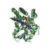
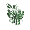
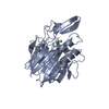
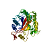
| Title | Crystal structure of beta toxin from Staphylococcus aureus F277A, P278A mutant with bound magnesium ions | ||||||
 Components Components | Beta-hemolysin | ||||||
 Keywords Keywords | TOXIN / beta toxin / hemolysin / sphingomyelinase | ||||||
| Function / homology |  Function and homology information Function and homology informationsphingomyelin phosphodiesterase activity / killing of cells of another organism / extracellular region / metal ion binding Similarity search - Function | ||||||
| Biological species |  Staphylococcus aureus RN4220 (bacteria) Staphylococcus aureus RN4220 (bacteria) | ||||||
| Method |  X-RAY DIFFRACTION / SYNCHROTRON / MOLECULAR REPLACEMENT / Resolution: 1.8 Å X-RAY DIFFRACTION / SYNCHROTRON / MOLECULAR REPLACEMENT / Resolution: 1.8 Å | ||||||
 Authors Authors | Huseby, M. / Shi, K. / Kruse, A.C. / Ohlendorf, D.H. | ||||||
 Citation Citation | Journal: to be published / Year: 2009 Title: Structure and biological functions of beta toxin from Staphylococcus aureus: Role of the hydrophobic beta hairpin in virulence Authors: Huseby, M. / Shi, K. / Kruse, A.C. / Digre, J. / Mengistu, F. / Bohach, G.A. / Schlievert, P.S. / Ohlendorf, D.H. / Earhart, C.A. #1: Journal: J.Bacteriol. / Year: 2007 Title: Structure and biological activities of beta toxin from Staphylococcus aureus. Authors: Huseby, M. / Shi, K. / Brown, C.K. / Digre, J. / Mengistu, F. / Seo, K.S. / Bohach, G.A. / Schlievert, P.M. / Ohlendorf, D.H. / Earhart, C.A. | ||||||
| History |
|
- Structure visualization
| Structure viewer | Molecule: MolmilJmol/JSmol |
|---|
- Downloads & links
-Download
| PDBx/mmCIF format | 3i48.cif.gz | 140 KB | Display | PDBx/mmCIF format |
|---|---|---|---|---|
| PDB format | pdb3i48.ent.gz | 107.5 KB | Display | PDB format |
| PDBx/mmJSON format | 3i48.json.gz | Tree view | PDBx/mmJSON format | |
| Others |  Other downloads Other downloads |
-Validation report
| Arichive directory | https://data.pdbj.org/pub/pdb/validation_reports/i4/3i48ftp://data.pdbj.org/pub/pdb/validation_reports/i4/3i48 | HTTPS FTP |
|---|
-Related structure data
| Related structure data |  3i41SC  3i46C  3i5vC S: Starting model for refinement C: citing same article ( |
|---|---|
| Similar structure data |


-Links
 PDBj
PDBj

- Assembly
| Deposited unit | 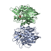
| ||||||||
|---|---|---|---|---|---|---|---|---|---|
| 1 | 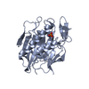
| ||||||||
| 2 | 
| ||||||||
| Unit cell |
|
-Components
| #1: Protein | Mass: 36092.156 Da / Num. of mol.: 2 / Fragment: UNP residues 35-330 / Mutation: F310A, P311A Source method: isolated from a genetically manipulated source Source: (gene. exp.) Staphylococcus aureus RN4220 (bacteria)Strain: RN2440 / Gene: hlb / Plasmid: pET28b / Production host: #2: Chemical |   Mass: 24.305 Da / Num. of mol.: 2 / Source method: obtained synthetically / Formula: Mg Mass: 24.305 Da / Num. of mol.: 2 / Source method: obtained synthetically / Formula: Mg#3: Chemical |   Mass: 94.971 Da / Num. of mol.: 2 / Source method: obtained synthetically / Formula: PO4 Mass: 94.971 Da / Num. of mol.: 2 / Source method: obtained synthetically / Formula: PO4#4: Water | ChemComp-HOH / |  Mass: 18.015 Da / Num. of mol.: 472 / Source method: isolated from a natural source / Formula: H2O Mass: 18.015 Da / Num. of mol.: 472 / Source method: isolated from a natural source / Formula: H2OHas protein modification | Y | |
|---|
-Experimental details
-Experiment
| Experiment | Method: X-RAY DIFFRACTION / Number of used crystals: 1 |
|---|
- Sample preparation
| Crystal | Density Matthews: 1.89 Å3/Da / Density % sol: 34.94 % |
|---|---|
| Crystal grow | Temperature: 291 K / Method: vapor diffusion, sitting drop / pH: 6.5 Details: PEG 4000, 0.1 M MES, 5mM MgCl2, pH 6.5, VAPOR DIFFUSION, SITTING DROP, temperature 291K |
-Data collection
| Diffraction | Mean temperature: 100 K |
|---|---|
| Diffraction source | Source: SYNCHROTRON / Site: APS  / Beamline: 14-BM-C / Wavelength: 0.9002 Å / Beamline: 14-BM-C / Wavelength: 0.9002 Å |
| Detector | Type: ADSC QUANTUM 315 / Detector: CCD / Date: Oct 11, 2007 / Details: mirrors |
| Radiation | Monochromator: bent Ge(111) / Protocol: SINGLE WAVELENGTH / Monochromatic (M) / Laue (L): M / Scattering type: x-ray |
| Radiation wavelength | Wavelength: 0.9002 Å / Relative weight: 1 |
| Reflection | Resolution: 1.8→50 Å / Num. all: 51464 / Num. obs: 51464 / % possible obs: 99.8 % / Observed criterion σ(I): 0 / Redundancy: 4.3 % / Rmerge(I) obs: 0.055 / Χ2: 1.387 / Net I/σ(I): 11.7 |
| Reflection shell | Resolution: 1.8→1.83 Å / Redundancy: 3.8 % / Rmerge(I) obs: 0.472 / Mean I/σ(I) obs: 2.6 / Num. unique all: 2493 / Χ2: 0.982 / % possible all: 99.5 |
- Processing
| Software |
| ||||||||||||||||||||||||||||||||||||||||||||||||||||||||||||||||||||||||||||||||||||||||||
|---|---|---|---|---|---|---|---|---|---|---|---|---|---|---|---|---|---|---|---|---|---|---|---|---|---|---|---|---|---|---|---|---|---|---|---|---|---|---|---|---|---|---|---|---|---|---|---|---|---|---|---|---|---|---|---|---|---|---|---|---|---|---|---|---|---|---|---|---|---|---|---|---|---|---|---|---|---|---|---|---|---|---|---|---|---|---|---|---|---|---|---|
| Refinement | Method to determine structure: MOLECULAR REPLACEMENT Starting model: PDB entry 3I41 Resolution: 1.8→28.16 Å / Cor.coef. Fo:Fc: 0.96 / Cor.coef. Fo:Fc free: 0.947 / Occupancy max: 1 / Occupancy min: 1 / Cross valid method: THROUGHOUT / ESU R: 0.144 / ESU R Free: 0.131 / Stereochemistry target values: MAXIMUM LIKELIHOOD / Details: HYDROGENS HAVE BEEN ADDED IN THE RIDING POSITIONS
| ||||||||||||||||||||||||||||||||||||||||||||||||||||||||||||||||||||||||||||||||||||||||||
| Solvent computation | Ion probe radii: 0.8 Å / Shrinkage radii: 0.8 Å / VDW probe radii: 1.2 Å / Solvent model: MASK | ||||||||||||||||||||||||||||||||||||||||||||||||||||||||||||||||||||||||||||||||||||||||||
| Displacement parameters | Biso max: 113.87 Å2 / Biso mean: 28.979 Å2 / Biso min: 13.34 Å2
| ||||||||||||||||||||||||||||||||||||||||||||||||||||||||||||||||||||||||||||||||||||||||||
| Refinement step | Cycle: LAST / Resolution: 1.8→28.16 Å
| ||||||||||||||||||||||||||||||||||||||||||||||||||||||||||||||||||||||||||||||||||||||||||
| Refine LS restraints |
| ||||||||||||||||||||||||||||||||||||||||||||||||||||||||||||||||||||||||||||||||||||||||||
| LS refinement shell | Resolution: 1.8→1.847 Å / Total num. of bins used: 20
|
