 Movie
Movie Controller


+ Open data

- Basic information
| Entry | Database: PDB / ID: 6kf7 | ||||||
|---|---|---|---|---|---|---|---|
| Title | Microbial Hormone-sensitive lipase E53 mutant S285G | ||||||
 Components Components | Lipase | ||||||
 Keywords Keywords | HYDROLASE / Esterase / Microbial Hormone-sensitive lipase | ||||||
| Function / homology |  Function and homology information Function and homology information | ||||||
| Biological species |  Erythrobacter longus (bacteria) Erythrobacter longus (bacteria) | ||||||
| Method |  X-RAY DIFFRACTION / SYNCHROTRON / MOLECULAR REPLACEMENT / Resolution: 1.8 Å X-RAY DIFFRACTION / SYNCHROTRON / MOLECULAR REPLACEMENT / Resolution: 1.8 Å | ||||||
 Authors Authors | Xiaochen, Y. / Zhengyang, L. / Xuewei, X. / Jixi, L. | ||||||
| Funding support |  China, 1items China, 1items
| ||||||
 Citation Citation | Journal: To Be Published Title: Microbial Hormone-sensitive lipase E53 mutant S285G Authors: Xiaochen, Y. | ||||||
| History |
|
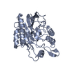
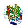
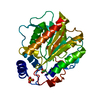
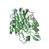
- Structure visualization
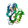
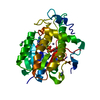
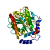
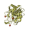
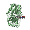
| Structure viewer | Molecule: MolmilJmol/JSmol |
|---|
- Downloads & links
-Download
| PDBx/mmCIF format | 6kf7.cif.gz | 338.8 KB | Display | PDBx/mmCIF format |
|---|---|---|---|---|
| PDB format | pdb6kf7.ent.gz | 219.8 KB | Display | PDB format |
| PDBx/mmJSON format | 6kf7.json.gz | Tree view | PDBx/mmJSON format | |
| Others |  Other downloads Other downloads |
-Validation report
| Arichive directory | https://data.pdbj.org/pub/pdb/validation_reports/kf/6kf7ftp://data.pdbj.org/pub/pdb/validation_reports/kf/6kf7 | HTTPS FTP |
|---|
-Related structure data
| Related structure data | 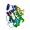 4ypvS S: Starting model for refinement |
|---|---|
| Similar structure data |

-Links
 PDBj
PDBj
- Assembly
| Deposited unit | 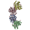
| ||||||||||
|---|---|---|---|---|---|---|---|---|---|---|---|
| 1 | 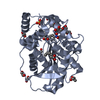
| ||||||||||
| 2 | 
| ||||||||||
| 3 | 
| ||||||||||
| 4 | 
| ||||||||||
| Unit cell |
| ||||||||||
| Components on special symmetry positions |
|
-Components
-Protein , 1 types, 4 molecules ABCD
| #1: Protein | Mass: 33128.703 Da / Num. of mol.: 4 / Mutation: S285G Source method: isolated from a genetically manipulated source Source: (gene. exp.) Erythrobacter longus (bacteria) / Gene: EH31_02760Production host: References: UniProt: A0A074MDU6 |
|---|
-Non-polymers , 8 types, 1448 molecules 


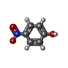

| #2: Chemical | ChemComp-D8F / (  Mass: 237.252 Da / Num. of mol.: 4 / Source method: obtained synthetically / Formula: C12H15NO4 / Feature type: SUBJECT OF INVESTIGATION Mass: 237.252 Da / Num. of mol.: 4 / Source method: obtained synthetically / Formula: C12H15NO4 / Feature type: SUBJECT OF INVESTIGATION#3: Chemical |  Mass: 96.063 Da / Num. of mol.: 2 / Source method: obtained synthetically / Formula: SO4 Mass: 96.063 Da / Num. of mol.: 2 / Source method: obtained synthetically / Formula: SO4#4: Chemical | ChemComp-EDO /  Mass: 62.068 Da / Num. of mol.: 13 / Source method: obtained synthetically / Formula: C2H6O2 Mass: 62.068 Da / Num. of mol.: 13 / Source method: obtained synthetically / Formula: C2H6O2#5: Chemical | ChemComp-GOL /  Mass: 92.094 Da / Num. of mol.: 16 / Source method: obtained synthetically / Formula: C3H8O3 Mass: 92.094 Da / Num. of mol.: 16 / Source method: obtained synthetically / Formula: C3H8O3#6: Chemical |  Mass: 116.158 Da / Num. of mol.: 2 / Source method: isolated from a natural source / Formula: C6H12O2 Mass: 116.158 Da / Num. of mol.: 2 / Source method: isolated from a natural source / Formula: C6H12O2#7: Chemical | ChemComp-PGE / |  Mass: 150.173 Da / Num. of mol.: 1 / Source method: obtained synthetically / Formula: C6H14O4 Mass: 150.173 Da / Num. of mol.: 1 / Source method: obtained synthetically / Formula: C6H14O4#8: Chemical | ChemComp-NPO / |  Mass: 139.109 Da / Num. of mol.: 1 / Source method: obtained synthetically / Formula: C6H5NO3 Mass: 139.109 Da / Num. of mol.: 1 / Source method: obtained synthetically / Formula: C6H5NO3#9: Water | ChemComp-HOH / | Mass: 18.015 Da / Num. of mol.: 1409 / Source method: isolated from a natural source / Formula: H2O |
|---|
-Details
| Has ligand of interest | Y |
|---|---|
| Has protein modification | N |
-Experimental details
-Experiment
| Experiment | Method: X-RAY DIFFRACTION / Number of used crystals: 1 |
|---|
- Sample preparation
| Crystal | Density Matthews: 3.8 Å3/Da / Density % sol: 67.62 % |
|---|---|
| Crystal grow | Temperature: 293 K / Method: vapor diffusion, hanging drop / Details: Calcium chloride, Bis-Tris, PEG MME 550, pH 6.5 |
-Data collection
| Diffraction | Mean temperature: 80 K / Serial crystal experiment: N |
|---|---|
| Diffraction source | Source: SYNCHROTRON / Site: SSRF / Beamline: BL19U1 / Wavelength: 0.97916 Å |
| Detector | Type: ADSC QUANTUM 315r / Detector: CCD / Date: May 1, 2017 |
| Radiation | Protocol: SINGLE WAVELENGTH / Monochromatic (M) / Laue (L): M / Scattering type: x-ray |
| Radiation wavelength | Wavelength: 0.97916 Å / Relative weight: 1 |
| Reflection | Resolution: 1.8→48.53 Å / Num. obs: 186934 / % possible obs: 99.62 % / Redundancy: 6.5 % / Biso Wilson estimate: 24.48 Å2 / Rmerge(I) obs: 0.119 / Net I/σ(I): 21.756 |
| Reflection shell | Resolution: 1.8→1.86 Å / Rmerge(I) obs: 0.548 / Num. unique obs: 18389 |
- Processing
| Software |
| |||||||||||||||||||||||||||||||||||||||||||||||||||||||||||||||||||||||||||||||||||||||||||||||||||||||||||||||||||||||||||||||||||||||||||||||||||||||||||||||||||||||||||||||||||||||||||||||||||||||||||||||||||||||||
|---|---|---|---|---|---|---|---|---|---|---|---|---|---|---|---|---|---|---|---|---|---|---|---|---|---|---|---|---|---|---|---|---|---|---|---|---|---|---|---|---|---|---|---|---|---|---|---|---|---|---|---|---|---|---|---|---|---|---|---|---|---|---|---|---|---|---|---|---|---|---|---|---|---|---|---|---|---|---|---|---|---|---|---|---|---|---|---|---|---|---|---|---|---|---|---|---|---|---|---|---|---|---|---|---|---|---|---|---|---|---|---|---|---|---|---|---|---|---|---|---|---|---|---|---|---|---|---|---|---|---|---|---|---|---|---|---|---|---|---|---|---|---|---|---|---|---|---|---|---|---|---|---|---|---|---|---|---|---|---|---|---|---|---|---|---|---|---|---|---|---|---|---|---|---|---|---|---|---|---|---|---|---|---|---|---|---|---|---|---|---|---|---|---|---|---|---|---|---|---|---|---|---|---|---|---|---|---|---|---|---|---|---|---|---|---|---|---|---|
| Refinement | Method to determine structure: MOLECULAR REPLACEMENT Starting model: 4YPV Resolution: 1.8→48.53 Å / SU ML: 0.1754 / Cross valid method: FREE R-VALUE / σ(F): 1.36 / Phase error: 18.9413
| |||||||||||||||||||||||||||||||||||||||||||||||||||||||||||||||||||||||||||||||||||||||||||||||||||||||||||||||||||||||||||||||||||||||||||||||||||||||||||||||||||||||||||||||||||||||||||||||||||||||||||||||||||||||||
| Solvent computation | Shrinkage radii: 0.9 Å / VDW probe radii: 1.11 Å | |||||||||||||||||||||||||||||||||||||||||||||||||||||||||||||||||||||||||||||||||||||||||||||||||||||||||||||||||||||||||||||||||||||||||||||||||||||||||||||||||||||||||||||||||||||||||||||||||||||||||||||||||||||||||
| Displacement parameters | Biso mean: 28.49 Å2 | |||||||||||||||||||||||||||||||||||||||||||||||||||||||||||||||||||||||||||||||||||||||||||||||||||||||||||||||||||||||||||||||||||||||||||||||||||||||||||||||||||||||||||||||||||||||||||||||||||||||||||||||||||||||||
| Refinement step | Cycle: LAST / Resolution: 1.8→48.53 Å
| |||||||||||||||||||||||||||||||||||||||||||||||||||||||||||||||||||||||||||||||||||||||||||||||||||||||||||||||||||||||||||||||||||||||||||||||||||||||||||||||||||||||||||||||||||||||||||||||||||||||||||||||||||||||||
| Refine LS restraints |
| |||||||||||||||||||||||||||||||||||||||||||||||||||||||||||||||||||||||||||||||||||||||||||||||||||||||||||||||||||||||||||||||||||||||||||||||||||||||||||||||||||||||||||||||||||||||||||||||||||||||||||||||||||||||||
| LS refinement shell |
|
