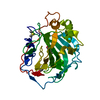
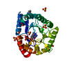
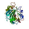
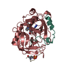
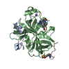
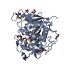
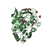
- PDB-4e5v: Crystal structure of a Putative thua-like protein (PARMER_02418) ... -
+
Open data
ID or keywords:
Loading...
-
Basic information
Entry
Database: PDB / ID: 4e5v
Title
Crystal structure of a Putative thua-like protein (PARMER_02418) from Parabacteroides merdae ATCC 43184 at 1.75 A resolution
Components
Putative thua-like protein
Keywords
STRUCTURAL GENOMICS / UNKNOWN FUNCTION / Thua-like proteins / trehalose utilisation / Joint Center for Structural Genomics / JCSG / Protein Structure Initiative / PSI-BIOLOGY
Function / homology
ThuA-like domain / Trehalose utilisation / Class I glutamine amidotransferase (GATase) domain / Class I glutamine amidotransferase-like / Rossmann fold / 3-Layer(aba) Sandwich / metal ion binding / Alpha Beta / ThuA domain-containing protein
Function and homology information
Biological species
Parabacteroides merdae (bacteria)
Method
X-RAY DIFFRACTION / SYNCHROTRON / MAD / Resolution: 1.75 Å
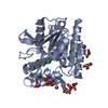
Mass: 18.015 Da / Num. of mol.: 641 / Source method: isolated from a natural source / Formula: H2O
Has protein modification
Y
Sequence details
THE CONSTRUCT WAS EXPRESSED WITH A PURIFICATION TAG MGSDKIHHHHHHENLYFQG. THE TAG WAS REMOVED WITH ...THE CONSTRUCT WAS EXPRESSED WITH A PURIFICATION TAG MGSDKIHHHHHHENLYFQG. THE TAG WAS REMOVED WITH TEV PROTEASE LEAVING ONLY A GLYCINE (0) FOLLOWED BY RESIDUES 25-304 OF THE TARGET SEQUENCE.
-
Experimental details
-
Experiment
Experiment
Method: X-RAY DIFFRACTION / Number of used crystals: 1
-
Sample preparation
Crystal
Density Matthews: 2.19 Å3/Da / Density % sol: 43.96 % Description: DATA WERE SCALED USING XSCALE WITH FRIEDEL PAIRS KEPT AS SEPARATE WHEN COMPUTING R-SYM, COMPLETENESS AND
Crystal grow
Temperature: 293 K / Method: vapor diffusion, sitting drop Details: 20.00% polyethylene glycol 3350, 0.200M ammonium fluoride, NANODROP, VAPOR DIFFUSION, SITTING DROP, temperature 293K
Type: DECTRIS PILATUS 6M / Detector: PIXEL / Date: Dec 7, 2011 Details: Flat mirror (vertical focusing); single crystal Si(111) bent monochromator (horizontal focusing)
Radiation
Monochromator: single crystal Si(111) bent / Protocol: MAD / Monochromatic (M) / Laue (L): M / Scattering type: x-ray
Radiation wavelength
ID
Wavelength (Å)
Relative weight
1
0.91837
1
2
0.97941
1
3
0.97882
1
Reflection
Resolution: 1.75→29.832 Å / Num. obs: 51578 / % possible obs: 81.3 % / Observed criterion σ(I): -3 / Biso Wilson estimate: 15.741 Å2 / Rmerge(I) obs: 0.066 / Net I/σ(I): 7.24
Reflection shell
Diffraction-ID: 1
Resolution (Å)
Highest resolution (Å)
Rmerge(I) obs
Mean I/σ(I) obs
Num. measured obs
Num. unique obs
% possible all
1.75-1.81
0.366
2
10084
8490
80.3
1.81-1.89
0.266
2.7
11248
9483
79
1.89-1.97
0.201
3.5
9177
7792
76.2
1.97-2.07
0.153
4.2
10455
8876
83.6
2.07-2.2
0.112
5.5
10858
9218
83.3
2.2-2.37
0.086
6.7
10690
9091
81.9
2.37-2.61
0.073
7.8
10276
8779
79.5
2.61-2.99
0.054
9.9
11107
9487
85.5
2.99-3.76
0.041
13
10072
8672
79.4
3.76
0.031
16.2
10932
9354
84.4
-
Phasing
Phasing
Method: MAD
-
Processing
Software
Name
Version
Classification
NB
MolProbity
3beta29
modelbuilding
PDB_EXTRACT
3.1
dataextraction
SHELX
phasing
SHARP
phasing
XSCALE
December6, 2010
datascaling
BUSTER-TNT
2.10.0
refinement
XDS
datareduction
SHELXD
phasing
BUSTER
2.10.0
refinement
Refinement
Method to determine structure: MAD / Resolution: 1.75→29.832 Å / Cor.coef. Fo:Fc: 0.9568 / Cor.coef. Fo:Fc free: 0.9436 / Occupancy max: 1 / Occupancy min: 0.4 / Cross valid method: THROUGHOUT / σ(F): 0 Details: 1. ATOM RECORD CONTAINS SUM OF TLS AND RESIDUAL B FACTORS. ANISOU RECORD CONTAINS SUM OF TLS AND RESIDUAL U FACTORS. 2. A MET-INHIBITION PROTOCOL WAS USED FOR SELENOMETHIONINE INCORPORATION ...Details: 1. ATOM RECORD CONTAINS SUM OF TLS AND RESIDUAL B FACTORS. ANISOU RECORD CONTAINS SUM OF TLS AND RESIDUAL U FACTORS. 2. A MET-INHIBITION PROTOCOL WAS USED FOR SELENOMETHIONINE INCORPORATION DURING PROTEIN EXPRESSION. THE OCCUPANCY OF THE SE ATOMS IN THE MSE RESIDUES WAS REDUCED TO 0.75 TO ACCOUNT FOR THE REDUCED SCATTERING POWER DUE TO PARTIAL S-MET INCORPORATION. 3. 1,2 ETHANEDIOL (EDO) FROM THE CRYOPROTECTANT AND FROM THE CRYSTALLIZATION CONDITION HAVE BEEN MODELED IN THE SOLVENT STRUCTURE. 4. ZINC IONS ARE MODELED IN THE STRUCTURE. THE ASSIGNMENT OF ZINC IS SUPPORTED BY COORDINATION GEOMETRY, ANOMALOUS DIFFERENCE FOURIER MAPS AND X-RAY FLUORESCENCE SCANS (WHICH SHOWED A PEAK FOR ZINC). ZINC WAS NOT ADDED DURING CRYSTALLIZATION OR PURIFICATION AND CO-PURIFIED WITH THE PROTEIN. 5. NCS RESTRAINTS WERE APPLIED USING BUSTER'S LSSR RESTRAINT REPRESENTATION (-AUTONCS). 6. THE MAD PHASES WERE USED AS RESTRAINTS DURING REFINEMENT.
In the structure databanks used in Yorodumi, some data are registered as the other names, "COVID-19 virus" and "2019-nCoV". Here are the details of the virus and the list of structure data.
Jan 31, 2019. EMDB accession codes are about to change! (news from PDBe EMDB page)
EMDB accession codes are about to change! (news from PDBe EMDB page)
The allocation of 4 digits for EMDB accession codes will soon come to an end. Whilst these codes will remain in use, new EMDB accession codes will include an additional digit and will expand incrementally as the available range of codes is exhausted. The current 4-digit format prefixed with “EMD-” (i.e. EMD-XXXX) will advance to a 5-digit format (i.e. EMD-XXXXX), and so on. It is currently estimated that the 4-digit codes will be depleted around Spring 2019, at which point the 5-digit format will come into force.
The EM Navigator/Yorodumi systems omit the EMD- prefix.
Related info.:Q: What is EMD? / ID/Accession-code notation in Yorodumi/EM Navigator
Yorodumi is a browser for structure data from EMDB, PDB, SASBDB, etc.
This page is also the successor to EM Navigator detail page, and also detail information page/front-end page for Omokage search.
The word "yorodu" (or yorozu) is an old Japanese word meaning "ten thousand". "mi" (miru) is to see.
Related info.:EMDB / PDB / SASBDB / Comparison of 3 databanks / Yorodumi Search / Aug 31, 2016. New EM Navigator & Yorodumi / Yorodumi Papers / Jmol/JSmol / Function and homology information / Changes in new EM Navigator and Yorodumi
 Movie
Movie Controller

 Yorodumi
 Open data
 Basic information
 Components
 Keywords
 Function and homology information
 Parabacteroides merdae (bacteria)
 X-RAY DIFFRACTION /
  Authors
 Citation
 Structure visualization
 Downloads & links
 Other downloads


 PDBj
PDBj

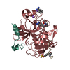
 Assembly
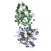

 Mass: 65.409 Da / Num. of mol.: 2 / Source method: obtained synthetically / Formula: Zn
 Mass: 62.068 Da / Num. of mol.: 16 / Source method: obtained synthetically / Formula: C2H6O2
 Mass: 18.015 Da / Num. of mol.: 641 / Source method: isolated from a natural source / Formula: H2O
 Sample preparation
 / Beamline: BL11-1 / Wavelength: 0.91837, 0.97941, 0.97882
 Processing