 Movie
Movie Controller

[English] 日本語
 Yorodumi
- PDB-4djz: Catalytic fragment of masp-1 in complex with its specific inhibit... -
+ Open data

- Basic information
| Entry | Database: PDB / ID: 4djz | ||||||
|---|---|---|---|---|---|---|---|
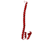
| Title | Catalytic fragment of masp-1 in complex with its specific inhibitor developed by directed evolution on sgci scaffold | ||||||
 Components Components |
| ||||||
 Keywords Keywords | HYDROLASE/HYDROLASE INHIBITOR / in vitro evolution / specific inhibitor / hydrolase / HYDROLASE-HYDROLASE INHIBITOR complex | ||||||
| Function / homology |  Function and homology information Function and homology informationFicolins bind to repetitive carbohydrate structures on the target cell surface / Lectin pathway of complement activation / negative regulation of complement activation / complement activation, lectin pathway / Scavenging by Class A Receptors / Initial triggering of complement / Hydrolases; Acting on peptide bonds (peptidases); Serine endopeptidases / serine-type endopeptidase inhibitor activity / calcium-dependent protein binding / peptidase activity ...Ficolins bind to repetitive carbohydrate structures on the target cell surface / Lectin pathway of complement activation / negative regulation of complement activation / complement activation, lectin pathway / Scavenging by Class A Receptors / Initial triggering of complement / Hydrolases; Acting on peptide bonds (peptidases); Serine endopeptidases / serine-type endopeptidase inhibitor activity / calcium-dependent protein binding / peptidase activity / serine-type endopeptidase activity / calcium ion binding / SARS-CoV-2 activates/modulates innate and adaptive immune responses / protein homodimerization activity / proteolysis / extracellular space / extracellular region / nucleoplasm / identical protein binding / cytosol Similarity search - Function | ||||||
| Biological species |  Homo sapiens (human) Homo sapiens (human) Schistocerca gregaria (desert locust) Schistocerca gregaria (desert locust) | ||||||
| Method |  X-RAY DIFFRACTION / SYNCHROTRON / MOLECULAR REPLACEMENT / Resolution: 3.2 Å X-RAY DIFFRACTION / SYNCHROTRON / MOLECULAR REPLACEMENT / Resolution: 3.2 Å | ||||||
 Authors Authors | Heja, D. / Harmat, V. / Fodor, K. / Wilmanns, M. / Dobo, J. / Kekesi, K.A. / Zavodszky, P. / Gal, P. / Pal, G. | ||||||
 Citation Citation | Journal: J.Biol.Chem. / Year: 2012 Title: Monospecific Inhibitors Show That Both Mannan-binding Lectin-associated Serine Protease-1 (MASP-1) and -2 Are Essential for Lectin Pathway Activation and Reveal Structural Plasticity of MASP-2. Authors: Heja, D. / Harmat, V. / Fodor, K. / Wilmanns, M. / Dobo, J. / Kekesi, K.A. / Zavodszky, P. / Gal, P. / Pal, G. | ||||||
| History |
|
- Structure visualization
| Structure viewer | Molecule: MolmilJmol/JSmol |
|---|
- Downloads & links
-Download
| PDBx/mmCIF format | 4djz.cif.gz | 337.3 KB | Display | PDBx/mmCIF format |
|---|---|---|---|---|
| PDB format | pdb4djz.ent.gz | 274.9 KB | Display | PDB format |
| PDBx/mmJSON format | 4djz.json.gz | Tree view | PDBx/mmJSON format | |
| Others |  Other downloads Other downloads |
-Validation report
| Arichive directory | https://data.pdbj.org/pub/pdb/validation_reports/dj/4djzftp://data.pdbj.org/pub/pdb/validation_reports/dj/4djz | HTTPS FTP |
|---|
-Related structure data
| Related structure data | 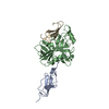 3tvjC  2xttS  3govS C: citing same article ( S: Starting model for refinement |
|---|---|
| Similar structure data |
-Links
 PDBj
PDBj


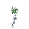
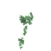
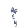
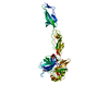
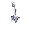
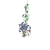
- Assembly
| Deposited unit | 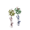
| |||||||||||||||||||||||||||||||||||||||||||||||||||||||||||||||||||||||||||||||||||||||||||||||||||||||||||||||||||||||||||||||||||||||||||||||||||||||||||||||||||||||||||||||||||||||||||||||||||||||
|---|---|---|---|---|---|---|---|---|---|---|---|---|---|---|---|---|---|---|---|---|---|---|---|---|---|---|---|---|---|---|---|---|---|---|---|---|---|---|---|---|---|---|---|---|---|---|---|---|---|---|---|---|---|---|---|---|---|---|---|---|---|---|---|---|---|---|---|---|---|---|---|---|---|---|---|---|---|---|---|---|---|---|---|---|---|---|---|---|---|---|---|---|---|---|---|---|---|---|---|---|---|---|---|---|---|---|---|---|---|---|---|---|---|---|---|---|---|---|---|---|---|---|---|---|---|---|---|---|---|---|---|---|---|---|---|---|---|---|---|---|---|---|---|---|---|---|---|---|---|---|---|---|---|---|---|---|---|---|---|---|---|---|---|---|---|---|---|---|---|---|---|---|---|---|---|---|---|---|---|---|---|---|---|---|---|---|---|---|---|---|---|---|---|---|---|---|---|---|---|---|
| 1 | 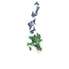
| |||||||||||||||||||||||||||||||||||||||||||||||||||||||||||||||||||||||||||||||||||||||||||||||||||||||||||||||||||||||||||||||||||||||||||||||||||||||||||||||||||||||||||||||||||||||||||||||||||||||
| 2 | 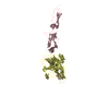
| |||||||||||||||||||||||||||||||||||||||||||||||||||||||||||||||||||||||||||||||||||||||||||||||||||||||||||||||||||||||||||||||||||||||||||||||||||||||||||||||||||||||||||||||||||||||||||||||||||||||
| Unit cell |
| |||||||||||||||||||||||||||||||||||||||||||||||||||||||||||||||||||||||||||||||||||||||||||||||||||||||||||||||||||||||||||||||||||||||||||||||||||||||||||||||||||||||||||||||||||||||||||||||||||||||
| Noncrystallographic symmetry (NCS) | NCS domain:
NCS domain segments:
|