 Movie
Movie Controller

[English] 日本語
 Yorodumi
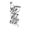
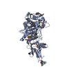
- PDB-3tvj: Catalytic fragment of MASP-2 in complex with its specific inhibit... -
+ Open data

- Basic information
| Entry | Database: PDB / ID: 3tvj | ||||||
|---|---|---|---|---|---|---|---|
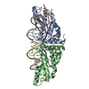
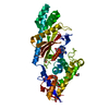
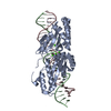
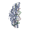
| Title | Catalytic fragment of MASP-2 in complex with its specific inhibitor developed by directed evolution on SGCI scaffold | ||||||
 Components Components |
| ||||||
 Keywords Keywords | HYDROLASE / in vitro evolution / specific inhibitor / allostery | ||||||
| Function / homology |  Function and homology information Function and homology informationmannan-binding lectin-associated serine protease-2 / complement component C4b binding / Ficolins bind to repetitive carbohydrate structures on the target cell surface / Lectin pathway of complement activation / complement activation, lectin pathway / Initial triggering of complement / complement activation, classical pathway / serine-type endopeptidase inhibitor activity / calcium-dependent protein binding / peptidase activity ...mannan-binding lectin-associated serine protease-2 / complement component C4b binding / Ficolins bind to repetitive carbohydrate structures on the target cell surface / Lectin pathway of complement activation / complement activation, lectin pathway / Initial triggering of complement / complement activation, classical pathway / serine-type endopeptidase inhibitor activity / calcium-dependent protein binding / peptidase activity / serine-type endopeptidase activity / calcium ion binding / SARS-CoV-2 activates/modulates innate and adaptive immune responses / proteolysis / : / extracellular exosome / extracellular region / identical protein binding Similarity search - Function | ||||||
| Biological species |  Homo sapiens (human) Homo sapiens (human) Schistocerca gregaria (desert locust) Schistocerca gregaria (desert locust) | ||||||
| Method |  X-RAY DIFFRACTION / SYNCHROTRON / MOLECULAR REPLACEMENT / Resolution: 1.28 Å X-RAY DIFFRACTION / SYNCHROTRON / MOLECULAR REPLACEMENT / Resolution: 1.28 Å | ||||||
 Authors Authors | Heja, D. / Harmat, V. / Dobo, J. / Szasz, R. / Kekesi, K.A. / Zavodszky, P. / Gal, P. / Pal, G. | ||||||
 Citation Citation | Journal: J.Biol.Chem. / Year: 2012 Title: Monospecific Inhibitors Show That Both Mannan-binding Lectin-associated Serine Protease-1 (MASP-1) and -2 Are Essential for Lectin Pathway Activation and Reveal Structural Plasticity of MASP-2. Authors: Heja, D. / Harmat, V. / Fodor, K. / Wilmanns, M. / Dobo, J. / Kekesi, K.A. / Zavodszky, P. / Gal, P. / Pal, G. | ||||||
| History |
|
- Structure visualization
| Structure viewer | Molecule: MolmilJmol/JSmol |
|---|
- Downloads & links
-Download
| PDBx/mmCIF format | 3tvj.cif.gz | 183.1 KB | Display | PDBx/mmCIF format |
|---|---|---|---|---|
| PDB format | pdb3tvj.ent.gz | 145.3 KB | Display | PDB format |
| PDBx/mmJSON format | 3tvj.json.gz | Tree view | PDBx/mmJSON format | |
| Others |  Other downloads Other downloads |
-Validation report
| Arichive directory | https://data.pdbj.org/pub/pdb/validation_reports/tv/3tvjftp://data.pdbj.org/pub/pdb/validation_reports/tv/3tvj | HTTPS FTP |
|---|
-Related structure data
| Related structure data |  4djzC  1gl1S  1q3xS C: citing same article ( S: Starting model for refinement |
|---|---|
| Similar structure data |


-Links
 PDBj
PDBj


- Assembly
| Deposited unit | 
| ||||||||
|---|---|---|---|---|---|---|---|---|---|
| 1 |
| ||||||||
| Unit cell |
|
-Components
| #1: Protein | Mass: 9217.346 Da / Num. of mol.: 1 / Fragment: Sushi 2 domain residues 363-444 Source method: isolated from a genetically manipulated source Source: (gene. exp.) Homo sapiens (human) / Gene: MASP2 / Plasmid: pET-17b / Production host:  References: UniProt: O00187, mannan-binding lectin-associated serine protease-2 |
|---|---|
| #2: Protein | Mass: 26542.971 Da / Num. of mol.: 1 / Fragment: Peptidase S1 domain residues 445-686 Source method: isolated from a genetically manipulated source Source: (gene. exp.) Homo sapiens (human) / Gene: MASP2 / Plasmid: pET-17b / Production host: References: UniProt: O00187, mannan-binding lectin-associated serine protease-2 |
| #3: Protein/peptide | Mass: 4020.530 Da / Num. of mol.: 1 / Mutation: A83V, L86K, K87L, A88W, P90N Source method: isolated from a genetically manipulated source Details: expressed as MBP fusion protein / Source: (gene. exp.) Schistocerca gregaria (desert locust) / Gene: GenBank: Y09605.1 / Plasmid: pMal-TEV-SGPI / Production host: |
| #4: Chemical | ChemComp-SO4 /   Mass: 96.063 Da / Num. of mol.: 1 / Source method: obtained synthetically / Formula: SO4 Mass: 96.063 Da / Num. of mol.: 1 / Source method: obtained synthetically / Formula: SO4 |
| #5: Water | ChemComp-HOH /  Mass: 18.015 Da / Num. of mol.: 509 / Source method: isolated from a natural source / Formula: H2O Mass: 18.015 Da / Num. of mol.: 509 / Source method: isolated from a natural source / Formula: H2O |
| Has protein modification | Y |
-Experimental details
-Experiment
| Experiment | Method: X-RAY DIFFRACTION / Number of used crystals: 1 |
|---|
- Sample preparation
| Crystal | Density Matthews: 2.28 Å3/Da / Density % sol: 46.11 % |
|---|---|
| Crystal grow | Temperature: 293 K / Method: vapor diffusion, hanging drop / pH: 6.5 Details: 24.5% PEG 5000 MME, 0.164 M ammonium sulphate, 82 mM MES, pH 6.5, VAPOR DIFFUSION, HANGING DROP, temperature 293K |
-Data collection
| Diffraction | Mean temperature: 100 K | |||||||||||||||||||||
|---|---|---|---|---|---|---|---|---|---|---|---|---|---|---|---|---|---|---|---|---|---|---|
| Diffraction source | Source: SYNCHROTRON / Site: ESRF  / Beamline: ID14-1 / Wavelength: 0.9334 Å / Beamline: ID14-1 / Wavelength: 0.9334 Å | |||||||||||||||||||||
| Detector | Type: ADSC QUANTUM 210 / Detector: CCD / Date: Apr 21, 2011 | |||||||||||||||||||||
| Radiation | Monochromator: Diamond (111), Ge(220) / Protocol: SINGLE WAVELENGTH / Monochromatic (M) / Laue (L): M / Scattering type: x-ray | |||||||||||||||||||||
| Radiation wavelength | Wavelength: 0.9334 Å / Relative weight: 1 | |||||||||||||||||||||
| Reflection | Resolution: 1.28→40 Å / Num. all: 90627 / Num. obs: 90627 / % possible obs: 95.8 % / Observed criterion σ(F): 0 / Observed criterion σ(I): -3 / Redundancy: 5.44 % / Biso Wilson estimate: 16.911 Å2 / Rmerge(I) obs: 0.065 / Net I/σ(I): 15.1 | |||||||||||||||||||||
| Reflection shell |
|
- Processing
| Software |
| ||||||||||||||||||||||||||||||||||||||||||||||||||||||||||||||||||||||||||||||||||||||||||||||||||||||||||||||||||||||||||||||||||||||||||||||||||||||||||||||||||||||||||
|---|---|---|---|---|---|---|---|---|---|---|---|---|---|---|---|---|---|---|---|---|---|---|---|---|---|---|---|---|---|---|---|---|---|---|---|---|---|---|---|---|---|---|---|---|---|---|---|---|---|---|---|---|---|---|---|---|---|---|---|---|---|---|---|---|---|---|---|---|---|---|---|---|---|---|---|---|---|---|---|---|---|---|---|---|---|---|---|---|---|---|---|---|---|---|---|---|---|---|---|---|---|---|---|---|---|---|---|---|---|---|---|---|---|---|---|---|---|---|---|---|---|---|---|---|---|---|---|---|---|---|---|---|---|---|---|---|---|---|---|---|---|---|---|---|---|---|---|---|---|---|---|---|---|---|---|---|---|---|---|---|---|---|---|---|---|---|---|---|---|---|---|
| Refinement | Method to determine structure: MOLECULAR REPLACEMENT Starting model: domains of PDB entry 1Q3X, and PDB entry 1GL1 Resolution: 1.28→38 Å / Cor.coef. Fo:Fc: 0.972 / Cor.coef. Fo:Fc free: 0.955 / SU B: 2.182 / SU ML: 0.041 / Cross valid method: THROUGHOUT / σ(F): 0 / ESU R: 0.052 / ESU R Free: 0.054 / Stereochemistry target values: MAXIMUM LIKELIHOOD / Details: HYDROGENS HAVE BEEN ADDED IN THE RIDING POSITIONS
| ||||||||||||||||||||||||||||||||||||||||||||||||||||||||||||||||||||||||||||||||||||||||||||||||||||||||||||||||||||||||||||||||||||||||||||||||||||||||||||||||||||||||||
| Solvent computation | Ion probe radii: 0.8 Å / Shrinkage radii: 0.8 Å / VDW probe radii: 1.4 Å / Solvent model: BABINET MODEL WITH MASK | ||||||||||||||||||||||||||||||||||||||||||||||||||||||||||||||||||||||||||||||||||||||||||||||||||||||||||||||||||||||||||||||||||||||||||||||||||||||||||||||||||||||||||
| Displacement parameters | Biso mean: 14.05 Å2
| ||||||||||||||||||||||||||||||||||||||||||||||||||||||||||||||||||||||||||||||||||||||||||||||||||||||||||||||||||||||||||||||||||||||||||||||||||||||||||||||||||||||||||
| Refinement step | Cycle: LAST / Resolution: 1.28→38 Å
| ||||||||||||||||||||||||||||||||||||||||||||||||||||||||||||||||||||||||||||||||||||||||||||||||||||||||||||||||||||||||||||||||||||||||||||||||||||||||||||||||||||||||||
| Refine LS restraints |
| ||||||||||||||||||||||||||||||||||||||||||||||||||||||||||||||||||||||||||||||||||||||||||||||||||||||||||||||||||||||||||||||||||||||||||||||||||||||||||||||||||||||||||
| LS refinement shell | Resolution: 1.28→1.313 Å / Total num. of bins used: 20
| ||||||||||||||||||||||||||||||||||||||||||||||||||||||||||||||||||||||||||||||||||||||||||||||||||||||||||||||||||||||||||||||||||||||||||||||||||||||||||||||||||||||||||
| Refinement TLS params. | Method: refined / Refine-ID: X-RAY DIFFRACTION
| ||||||||||||||||||||||||||||||||||||||||||||||||||||||||||||||||||||||||||||||||||||||||||||||||||||||||||||||||||||||||||||||||||||||||||||||||||||||||||||||||||||||||||
| Refinement TLS group |
|
