 Movie
Movie Controller


+ Open data

- Basic information
| Entry | Database: PDB / ID: 3fd2 | ||||||
|---|---|---|---|---|---|---|---|
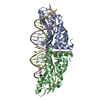
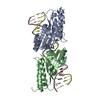
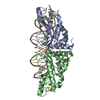
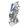
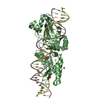
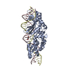
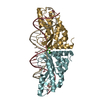
| Title | Crystal structure of mMsoI/DNA complex with calcium | ||||||
 Components Components |
| ||||||
 Keywords Keywords | HYDROLASE/DNA / Protein-DNA complex / Chloroplast / HYDROLASE-DNA COMPLEX | ||||||
| Function / homology |  Function and homology information Function and homology information | ||||||
| Biological species |  Monomastix sp. (plant) Monomastix sp. (plant)Monomastix sp. OKE-1 unidentified (others) synthetic construct (others) | ||||||
| Method |  X-RAY DIFFRACTION / MOLECULAR REPLACEMENT / Resolution: 2.69 Å X-RAY DIFFRACTION / MOLECULAR REPLACEMENT / Resolution: 2.69 Å | ||||||
 Authors Authors | Li, H. / Monnat, R.J. | ||||||
 Citation Citation | Journal: Nucleic Acids Res. / Year: 2009 Title: Generation of single-chain LAGLIDADG homing endonucleases from native homodimeric precursor proteins. Authors: Li, H. / Pellenz, S. / Ulge, U. / Stoddard, B.L. / Monnat, R.J. | ||||||
| History |
|
- Structure visualization
| Structure viewer | Molecule: MolmilJmol/JSmol |
|---|
- Downloads & links
-Download
| PDBx/mmCIF format | 3fd2.cif.gz | 110 KB | Display | PDBx/mmCIF format |
|---|---|---|---|---|
| PDB format | pdb3fd2.ent.gz | 79.1 KB | Display | PDB format |
| PDBx/mmJSON format | 3fd2.json.gz | Tree view | PDBx/mmJSON format | |
| Others |  Other downloads Other downloads |
-Validation report
| Arichive directory | https://data.pdbj.org/pub/pdb/validation_reports/fd/3fd2ftp://data.pdbj.org/pub/pdb/validation_reports/fd/3fd2 | HTTPS FTP |
|---|
-Related structure data
| Related structure data | 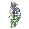 1m5xS S: Starting model for refinement |
|---|---|
| Similar structure data |


-Links
 PDBj
PDBj


- Assembly
| Deposited unit | 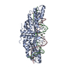
| ||||||||
|---|---|---|---|---|---|---|---|---|---|
| 1 |
| ||||||||
| Unit cell |
|
-Components
| #1: Protein | Mass: 42024.996 Da / Num. of mol.: 1 Source method: isolated from a genetically manipulated source Details: fusion protein comprises residues 1-170 of I-MSOI, a linker of 33 residues (TGSGSGSKHPTLTLPTTTSQENLPNGSGSGSGT) and a 2nd I-MSOI (1-170 residues). Source: (gene. exp.) Monomastix sp. (strain OKE-1) (plant), (gene. exp.) Monomastix sp. OKE-1, unidentifiedStrain: OKE-1 Gene: orf170, the chloroplast large subunit rDNA intron gene Plasmid: pET system / Production host:  References: UniProt: C0JWR6, Hydrolases; Acting on ester bonds | ||||
|---|---|---|---|---|---|
| #2: DNA chain | Mass: 7419.792 Da / Num. of mol.: 1 / Source method: obtained synthetically Details: the physiological DNA target site of I-MsoI in the chloroplast large subunit rDNA of Monomastix Source: (synth.) synthetic construct (others) | ||||
| #3: DNA chain | Mass: 7321.716 Da / Num. of mol.: 1 / Source method: obtained synthetically Details: the physiological DNA target site of I-MsoI in the chloroplast large subunit rDNA of Monomastix Source: (synth.) synthetic construct (others) | ||||
| #4: Chemical |   Mass: 40.078 Da / Num. of mol.: 2 / Source method: obtained synthetically / Formula: Ca Mass: 40.078 Da / Num. of mol.: 2 / Source method: obtained synthetically / Formula: Ca#5: Water | ChemComp-HOH / |  Mass: 18.015 Da / Num. of mol.: 35 / Source method: isolated from a natural source / Formula: H2O Mass: 18.015 Da / Num. of mol.: 35 / Source method: isolated from a natural source / Formula: H2OSequence details | 33 RESIDUES OF LINKER (TGSGSGSKHP | |
-Experimental details
-Experiment
| Experiment | Method: X-RAY DIFFRACTION / Number of used crystals: 1 |
|---|
- Sample preparation
| Crystal | Density Matthews: 1.93 Å3/Da / Density % sol: 36.4 % |
|---|---|
| Crystal grow | Temperature: 291 K / pH: 7.3 Details: 22% PEG 400, 100 mM Tris pH 7.3, 10 mM calcium chloride, 20 mM sodium chloride, VAPOR DIFFUSION, HANGING DROP, temperature 291K |
-Data collection
| Diffraction source | Source: ROTATING ANODE / Type: OTHER / Wavelength: 1.54 |
|---|---|
| Detector | Type: RIGAKU RAXIS IV / Detector: IMAGE PLATE / Date: Mar 25, 2008 |
| Radiation | Protocol: SINGLE WAVELENGTH / Monochromatic (M) / Laue (L): M / Scattering type: x-ray |
| Radiation wavelength | Wavelength: 1.54 Å / Relative weight: 1 |
| Reflection | Resolution: 2.69→50 Å / Num. obs: 11073 / % possible obs: 97 % / Redundancy: 3.9 % / Rmerge(I) obs: 0.052 / Net I/σ(I): 21 |
| Reflection shell | Resolution: 2.69→2.8 Å / Redundancy: 3.9 % / Rmerge(I) obs: 0.258 / Mean I/σ(I) obs: 4.51 / % possible all: 95.8 |
- Processing
| Software |
| ||||||||||||||||||||||||||||||||||||||||||||||||||||||||||||||||||||||||||||||||||||||||||||||||||||||
|---|---|---|---|---|---|---|---|---|---|---|---|---|---|---|---|---|---|---|---|---|---|---|---|---|---|---|---|---|---|---|---|---|---|---|---|---|---|---|---|---|---|---|---|---|---|---|---|---|---|---|---|---|---|---|---|---|---|---|---|---|---|---|---|---|---|---|---|---|---|---|---|---|---|---|---|---|---|---|---|---|---|---|---|---|---|---|---|---|---|---|---|---|---|---|---|---|---|---|---|---|---|---|---|
| Refinement | Method to determine structure: MOLECULAR REPLACEMENT Starting model: 1M5X Resolution: 2.69→50 Å / Cor.coef. Fo:Fc: 0.953 / Cor.coef. Fo:Fc free: 0.901 / SU ML: 0.422 / TLS residual ADP flag: LIKELY RESIDUAL / Cross valid method: THROUGHOUT / ESU R Free: 0.457 / Stereochemistry target values: MAXIMUM LIKELIHOOD / Details: HYDROGENS HAVE BEEN ADDED IN THE RIDING POSITIONS
| ||||||||||||||||||||||||||||||||||||||||||||||||||||||||||||||||||||||||||||||||||||||||||||||||||||||
| Solvent computation | Ion probe radii: 0.8 Å / Shrinkage radii: 0.8 Å / VDW probe radii: 1.2 Å / Solvent model: MASK | ||||||||||||||||||||||||||||||||||||||||||||||||||||||||||||||||||||||||||||||||||||||||||||||||||||||
| Displacement parameters | Biso mean: 59.2 Å2
| ||||||||||||||||||||||||||||||||||||||||||||||||||||||||||||||||||||||||||||||||||||||||||||||||||||||
| Refinement step | Cycle: LAST / Resolution: 2.69→50 Å
| ||||||||||||||||||||||||||||||||||||||||||||||||||||||||||||||||||||||||||||||||||||||||||||||||||||||
| Refine LS restraints |
| ||||||||||||||||||||||||||||||||||||||||||||||||||||||||||||||||||||||||||||||||||||||||||||||||||||||
| LS refinement shell | Resolution: 2.69→2.76 Å / Total num. of bins used: 20
| ||||||||||||||||||||||||||||||||||||||||||||||||||||||||||||||||||||||||||||||||||||||||||||||||||||||
| Refinement TLS params. | Method: refined / Refine-ID: X-RAY DIFFRACTION
| ||||||||||||||||||||||||||||||||||||||||||||||||||||||||||||||||||||||||||||||||||||||||||||||||||||||
| Refinement TLS group |
|
