 Movie
Movie Controller

[English] 日本語
 Yorodumi
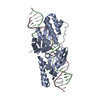
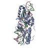
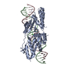
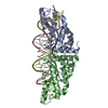
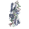
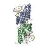
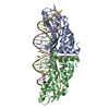
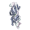
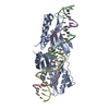
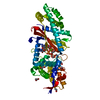
- PDB-5t2n: Engineered variant of I-OnuI meganuclease targeting the Anopheles... -
+ Open data

- Basic information
| Entry | Database: PDB / ID: 5t2n | ||||||||||||
|---|---|---|---|---|---|---|---|---|---|---|---|---|---|
| Title | Engineered variant of I-OnuI meganuclease targeting the Anopheles AGAP007280 gene; harbors 38 point mutations relative to wild-type I-OnuI | ||||||||||||
 Components Components |
| ||||||||||||
 Keywords Keywords | Hydrolase/DNA / Meganuclease / Engineered protein / DNA complex / Homing Endonuclease / Hydrolase-DNA complex | ||||||||||||
| Function / homology | Homing endonucleases / Endonuclease I-creI / Roll / Alpha Beta / DNA / DNA (> 10) Function and homology information Function and homology information | ||||||||||||
| Biological species | synthetic construct (others) | ||||||||||||
| Method |  X-RAY DIFFRACTION / SYNCHROTRON / MOLECULAR REPLACEMENT / molecular replacement / Resolution: 2.079 Å X-RAY DIFFRACTION / SYNCHROTRON / MOLECULAR REPLACEMENT / molecular replacement / Resolution: 2.079 Å | ||||||||||||
 Authors Authors | Stoddard, B.L. / Werther, R. | ||||||||||||
| Funding support |  United States, 3items United States, 3items
| ||||||||||||
 Citation Citation | Journal: Nucleic Acids Res. / Year: 2017 Title: Crystallographic analyses illustrate significant plasticity and efficient recoding of meganuclease target specificity. Authors: Werther, R. / Hallinan, J.P. / Lambert, A.R. / Havens, K. / Pogson, M. / Jarjour, J. / Galizi, R. / Windbichler, N. / Crisanti, A. / Nolan, T. / Stoddard, B.L. | ||||||||||||
| History |
|
- Structure visualization
| Structure viewer | Molecule: MolmilJmol/JSmol |
|---|
- Downloads & links
-Download
| PDBx/mmCIF format | 5t2n.cif.gz | 106.3 KB | Display | PDBx/mmCIF format |
|---|---|---|---|---|
| PDB format | pdb5t2n.ent.gz | 73.9 KB | Display | PDB format |
| PDBx/mmJSON format | 5t2n.json.gz | Tree view | PDBx/mmJSON format | |
| Others |  Other downloads Other downloads |
-Validation report
| Arichive directory | https://data.pdbj.org/pub/pdb/validation_reports/t2/5t2nftp://data.pdbj.org/pub/pdb/validation_reports/t2/5t2n | HTTPS FTP |
|---|
-Related structure data
| Related structure data |  5t2hC  5t2oC  3qqyS  5t2j C: citing same article ( S: Starting model for refinement |
|---|---|
| Similar structure data |
-Links
 PDBj
PDBj


- Assembly
| Deposited unit | 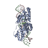
| ||||||||
|---|---|---|---|---|---|---|---|---|---|
| 1 |
| ||||||||
| Unit cell |
|
-Components
| #1: Protein | Mass: 34192.773 Da / Num. of mol.: 1 Source method: isolated from a genetically manipulated source Source: (gene. exp.) synthetic construct (others) / Production host:  | ||
|---|---|---|---|
| #2: DNA chain | Mass: 7802.002 Da / Num. of mol.: 1 / Source method: obtained synthetically / Source: (synth.) synthetic construct (others) | ||
| #3: DNA chain | Mass: 8176.281 Da / Num. of mol.: 1 / Source method: obtained synthetically / Source: (synth.) synthetic construct (others) | ||
| #4: Chemical | ChemComp-CA /   Mass: 40.078 Da / Num. of mol.: 4 / Source method: obtained synthetically / Formula: Ca Mass: 40.078 Da / Num. of mol.: 4 / Source method: obtained synthetically / Formula: Ca#5: Water | ChemComp-HOH / |  Mass: 18.015 Da / Num. of mol.: 59 / Source method: isolated from a natural source / Formula: H2O Mass: 18.015 Da / Num. of mol.: 59 / Source method: isolated from a natural source / Formula: H2O |
-Experimental details
-Experiment
| Experiment | Method: X-RAY DIFFRACTION / Number of used crystals: 1 |
|---|
- Sample preparation
| Crystal | Density Matthews: 2.29 Å3/Da / Density % sol: 46.27 % |
|---|---|
| Crystal grow | Temperature: 293 K / Method: vapor diffusion, hanging drop / pH: 6.5 Details: 0.05M ammonium sulfate, 0.05M BIS-TRIS pH 6.5, 35% pentaerythritol ethoxylate (15/4 EO/OH) |
-Data collection
| Diffraction | Mean temperature: 108 K | |||||||||||||||||||||||||||||||||||||||||||||||||||||||||||||||||||||||||||||||||||||||||||||||||||
|---|---|---|---|---|---|---|---|---|---|---|---|---|---|---|---|---|---|---|---|---|---|---|---|---|---|---|---|---|---|---|---|---|---|---|---|---|---|---|---|---|---|---|---|---|---|---|---|---|---|---|---|---|---|---|---|---|---|---|---|---|---|---|---|---|---|---|---|---|---|---|---|---|---|---|---|---|---|---|---|---|---|---|---|---|---|---|---|---|---|---|---|---|---|---|---|---|---|---|---|---|
| Diffraction source | Source: SYNCHROTRON / Site: ALS / Beamline: 5.0.2 / Wavelength: 1.084 Å | |||||||||||||||||||||||||||||||||||||||||||||||||||||||||||||||||||||||||||||||||||||||||||||||||||
| Detector | Type: DECTRIS PILATUS3 S 6M / Detector: PIXEL / Date: May 26, 2016 | |||||||||||||||||||||||||||||||||||||||||||||||||||||||||||||||||||||||||||||||||||||||||||||||||||
| Radiation | Protocol: SINGLE WAVELENGTH / Monochromatic (M) / Laue (L): M / Scattering type: x-ray | |||||||||||||||||||||||||||||||||||||||||||||||||||||||||||||||||||||||||||||||||||||||||||||||||||
| Radiation wavelength | Wavelength: 1.084 Å / Relative weight: 1 | |||||||||||||||||||||||||||||||||||||||||||||||||||||||||||||||||||||||||||||||||||||||||||||||||||
| Reflection | Resolution: 2.08→50 Å / Num. obs: 28554 / % possible obs: 99.6 % / Redundancy: 11.9 % / Biso Wilson estimate: 49.74 Å2 / Rmerge(I) obs: 0.094 / Rpim(I) all: 0.028 / Rrim(I) all: 0.098 / Χ2: 0.824 / Net I/av σ(I): 19.082 / Net I/σ(I): 10.1 / Num. measured all: 339574 | |||||||||||||||||||||||||||||||||||||||||||||||||||||||||||||||||||||||||||||||||||||||||||||||||||
| Reflection shell | Diffraction-ID: 1 / Rejects: _
|
-Phasing
| Phasing | Method: molecular replacement | |||||||||
|---|---|---|---|---|---|---|---|---|---|---|
| Phasing MR |
|
- Processing
| Software |
| |||||||||||||||||||||||||||||||||||||||||||||||||||||||||||||||||||||||||||||||||||||||||||||||||||||||||
|---|---|---|---|---|---|---|---|---|---|---|---|---|---|---|---|---|---|---|---|---|---|---|---|---|---|---|---|---|---|---|---|---|---|---|---|---|---|---|---|---|---|---|---|---|---|---|---|---|---|---|---|---|---|---|---|---|---|---|---|---|---|---|---|---|---|---|---|---|---|---|---|---|---|---|---|---|---|---|---|---|---|---|---|---|---|---|---|---|---|---|---|---|---|---|---|---|---|---|---|---|---|---|---|---|---|---|
| Refinement | Method to determine structure: MOLECULAR REPLACEMENT Starting model: 3QQY Resolution: 2.079→42.908 Å / SU ML: 0.29 / Cross valid method: FREE R-VALUE / σ(F): 1.34 / Phase error: 30.66 / Stereochemistry target values: ML
| |||||||||||||||||||||||||||||||||||||||||||||||||||||||||||||||||||||||||||||||||||||||||||||||||||||||||
| Solvent computation | Shrinkage radii: 0.9 Å / VDW probe radii: 1.11 Å / Solvent model: FLAT BULK SOLVENT MODEL | |||||||||||||||||||||||||||||||||||||||||||||||||||||||||||||||||||||||||||||||||||||||||||||||||||||||||
| Displacement parameters | Biso max: 111.61 Å2 / Biso mean: 56.1879 Å2 / Biso min: 31.27 Å2 | |||||||||||||||||||||||||||||||||||||||||||||||||||||||||||||||||||||||||||||||||||||||||||||||||||||||||
| Refinement step | Cycle: final / Resolution: 2.079→42.908 Å
| |||||||||||||||||||||||||||||||||||||||||||||||||||||||||||||||||||||||||||||||||||||||||||||||||||||||||
| Refine LS restraints |
| |||||||||||||||||||||||||||||||||||||||||||||||||||||||||||||||||||||||||||||||||||||||||||||||||||||||||
| LS refinement shell | Refine-ID: X-RAY DIFFRACTION / Total num. of bins used: 14
|
