 Movie
Movie Controller

[English] 日本語
 Yorodumi
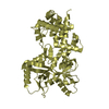
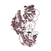
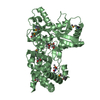
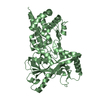
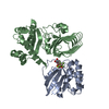
- PDB-4dgk: Crystal structure of Phytoene desaturase CRTI from Pantoea ananatis -
+ Open data

- Basic information
| Entry | Database: PDB / ID: 4dgk | ||||||
|---|---|---|---|---|---|---|---|
| Title | Crystal structure of Phytoene desaturase CRTI from Pantoea ananatis | ||||||
 Components Components | Phytoene dehydrogenase | ||||||
 Keywords Keywords | OXIDOREDUCTASE / the FAD/NAD(P)-binding Rossmann fold | ||||||
| Function / homology |  Function and homology information Function and homology informationphytoene desaturase (lycopene-forming) / carotene biosynthetic process / carotenoid biosynthetic process / oxidoreductase activity, acting on the CH-CH group of donors / FAD binding / plasma membrane Similarity search - Function | ||||||
| Biological species |  Pantoea ananatis (bacteria) Pantoea ananatis (bacteria) | ||||||
| Method |  X-RAY DIFFRACTION / SYNCHROTRON / MAD / Resolution: 2.35 Å X-RAY DIFFRACTION / SYNCHROTRON / MAD / Resolution: 2.35 Å | ||||||
 Authors Authors | Schaub, P. / Yu, Q. / Gemmecker, S. / Poussin-Courmontagne, P. / Mailliot, J. / McEwen, A.G. / Ghisla, S. / Beyer, P. / Cavarelli, J. | ||||||
 Citation Citation | Journal: Plos One / Year: 2012 Title: On the structure and function of the phytoene desaturase CRTI from Pantoea ananatis, a membrane-peripheral and FAD-dependent oxidase/isomerase. Authors: Schaub, P. / Yu, Q. / Gemmecker, S. / Poussin-Courmontagne, P. / Mailliot, J. / McEwen, A.G. / Ghisla, S. / Al-Babili, S. / Cavarelli, J. / Beyer, P. | ||||||
| History |
|
- Structure visualization
| Structure viewer | Molecule: MolmilJmol/JSmol |
|---|
- Downloads & links
-Download
| PDBx/mmCIF format | 4dgk.cif.gz | 176.8 KB | Display | PDBx/mmCIF format |
|---|---|---|---|---|
| PDB format | pdb4dgk.ent.gz | 141.1 KB | Display | PDB format |
| PDBx/mmJSON format | 4dgk.json.gz | Tree view | PDBx/mmJSON format | |
| Others |  Other downloads Other downloads |
-Validation report
| Summary document | 4dgk_validation.pdf.gz | 447.2 KB | Display | wwPDB validaton report |
|---|---|---|---|---|
| Full document | 4dgk_full_validation.pdf.gz | 452.1 KB | Display | |
| Data in XML | 4dgk_validation.xml.gz | 16.6 KB | Display | |
| Data in CIF | 4dgk_validation.cif.gz | 22.6 KB | Display | |
| Arichive directory | https://data.pdbj.org/pub/pdb/validation_reports/dg/4dgkftp://data.pdbj.org/pub/pdb/validation_reports/dg/4dgk | HTTPS FTP |
-Related structure data
| Similar structure data |
|---|


-Links
 PDBj
PDBj
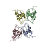
- Assembly
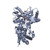
| Deposited unit | 
| ||||||||
|---|---|---|---|---|---|---|---|---|---|
| 1 |
| ||||||||
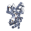
| Unit cell |
|
-Components
| #1: Protein | Mass: 56102.391 Da / Num. of mol.: 1 Source method: isolated from a genetically manipulated source Source: (gene. exp.) Pantoea ananatis (bacteria) / Gene: crtI / Production host: References: UniProt: P21685, Oxidoreductases; Acting on paired donors, with incorporation or reduction of molecular oxygen; Miscellaneous | ||||
|---|---|---|---|---|---|
| #2: Chemical | ChemComp-EDO /   Mass: 62.068 Da / Num. of mol.: 5 / Source method: obtained synthetically / Formula: C2H6O2 Mass: 62.068 Da / Num. of mol.: 5 / Source method: obtained synthetically / Formula: C2H6O2#3: Chemical | ChemComp-CL / |   Mass: 35.453 Da / Num. of mol.: 1 / Source method: obtained synthetically / Formula: Cl Mass: 35.453 Da / Num. of mol.: 1 / Source method: obtained synthetically / Formula: Cl#4: Water | ChemComp-HOH / |  Mass: 18.015 Da / Num. of mol.: 67 / Source method: isolated from a natural source / Formula: H2O Mass: 18.015 Da / Num. of mol.: 67 / Source method: isolated from a natural source / Formula: H2O |
-Experimental details
-Experiment
| Experiment | Method: X-RAY DIFFRACTION / Number of used crystals: 1 |
|---|
- Sample preparation
| Crystal | Density Matthews: 2.75 Å3/Da / Density % sol: 55.3 % |
|---|---|
| Crystal grow | Temperature: 290 K / pH: 6.2 Details: 8% PEG 8K, 0.1 M NaCl, 0.1 M Na/K phosphate pH 6.2, VAPOR DIFFUSION, temperature 290K |
-Data collection
| Diffraction | Mean temperature: 100 K | ||||||||||||
|---|---|---|---|---|---|---|---|---|---|---|---|---|---|
| Diffraction source | Source: SYNCHROTRON / Site: SOLEIL  / Beamline: PROXIMA 1 / Wavelength: 0.9791, 0.9794, 0.9770 / Beamline: PROXIMA 1 / Wavelength: 0.9791, 0.9794, 0.9770 | ||||||||||||
| Detector | Type: ADSC QUANTUM 315r / Detector: CCD / Date: Mar 18, 2009 | ||||||||||||
| Radiation | Monochromator: CHANNEL CUT CRYOGENICALLY COOLED MONOCHROMATOR CRYSTAL Protocol: MAD / Monochromatic (M) / Laue (L): M / Scattering type: x-ray | ||||||||||||
| Radiation wavelength |
| ||||||||||||
| Reflection | Resolution: 2.35→27.85 Å / Num. obs: 26274 / % possible obs: 99.8 % / Observed criterion σ(I): 0 / Redundancy: 6.5 % / Biso Wilson estimate: 68.8 Å2 / Rmerge(I) obs: 0.056 / Rsym value: 0.056 / Net I/σ(I): 22.5 | ||||||||||||
| Reflection shell | Resolution: 2.35→2.39 Å / Redundancy: 3.8 % / Rmerge(I) obs: 0.353 / Mean I/σ(I) obs: 4.4 / Rsym value: 0.353 / % possible all: 99.8 |
- Processing
| Software |
| ||||||||||||||||||||||||||||||||||||||||||||||||||||||||||||||||||||||||||||||||||||||||||||||||||||||||||||||||||
|---|---|---|---|---|---|---|---|---|---|---|---|---|---|---|---|---|---|---|---|---|---|---|---|---|---|---|---|---|---|---|---|---|---|---|---|---|---|---|---|---|---|---|---|---|---|---|---|---|---|---|---|---|---|---|---|---|---|---|---|---|---|---|---|---|---|---|---|---|---|---|---|---|---|---|---|---|---|---|---|---|---|---|---|---|---|---|---|---|---|---|---|---|---|---|---|---|---|---|---|---|---|---|---|---|---|---|---|---|---|---|---|---|---|---|---|
| Refinement | Method to determine structure: MAD / Resolution: 2.35→27.85 Å / Cor.coef. Fo:Fc: 0.95 / Cor.coef. Fo:Fc free: 0.923 / SU R Cruickshank DPI: 0.223 / Cross valid method: THROUGHOUT / σ(F): 0 / Stereochemistry target values: Engh & Huber Details: TLS SELECTION GROUP 1 { A|1 - A|33 A|59 - A|78 A|218 - A|275 A|456 - A|492 } GROUP 2 { A|41 - A|58 A|79 - A|100 A|212 - A|217 A|303 - A|425 } GROUP 3 { A|101 - A|211 }
| ||||||||||||||||||||||||||||||||||||||||||||||||||||||||||||||||||||||||||||||||||||||||||||||||||||||||||||||||||
| Displacement parameters | Biso mean: 72.87 Å2
| ||||||||||||||||||||||||||||||||||||||||||||||||||||||||||||||||||||||||||||||||||||||||||||||||||||||||||||||||||
| Refine analyze | Luzzati coordinate error obs: 0.39 Å | ||||||||||||||||||||||||||||||||||||||||||||||||||||||||||||||||||||||||||||||||||||||||||||||||||||||||||||||||||
| Refinement step | Cycle: LAST / Resolution: 2.35→27.85 Å
| ||||||||||||||||||||||||||||||||||||||||||||||||||||||||||||||||||||||||||||||||||||||||||||||||||||||||||||||||||
| Refine LS restraints |
| ||||||||||||||||||||||||||||||||||||||||||||||||||||||||||||||||||||||||||||||||||||||||||||||||||||||||||||||||||
| LS refinement shell | Resolution: 2.35→2.45 Å / Total num. of bins used: 13
| ||||||||||||||||||||||||||||||||||||||||||||||||||||||||||||||||||||||||||||||||||||||||||||||||||||||||||||||||||
| Refinement TLS params. | Method: refined / Refine-ID: X-RAY DIFFRACTION
| ||||||||||||||||||||||||||||||||||||||||||||||||||||||||||||||||||||||||||||||||||||||||||||||||||||||||||||||||||
| Refinement TLS group |
|
