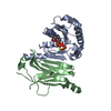
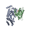
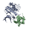
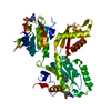
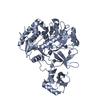
- PDB-3bh7: Crystal structure of the RP2-Arl3 complex bound to GDP-AlF4 -
+
Open data
ID or keywords:
Loading...
-
Basic information
Entry
Database: PDB / ID: 3bh7
Title
Crystal structure of the RP2-Arl3 complex bound to GDP-AlF4
Components
ADP-ribosylation factor-like protein 3
Protein XRP2
Keywords
SIGNALING PROTEIN / Protein-Protein complex / GTPase Activating protein and GTPase / Retinitis pigmentosa / GTP-binding / Lipoprotein / Myristate / Nucleotide-binding / Disease mutation / Membrane / Palmitate / Phosphoprotein / Sensory transduction / Vision / METAL BINDING PROTEIN
Function / homology
Function and homology information
Trafficking of myristoylated proteins to the cilium / Trafficking of myristoylated proteins to the cilium / photoreceptor cell development / periciliary membrane compartment / protein localization to ciliary membrane / intraciliary transport / post-Golgi vesicle-mediated transport / photoreceptor connecting cilium / protein localization to cilium / Golgi to plasma membrane transport ...Trafficking of myristoylated proteins to the cilium / Trafficking of myristoylated proteins to the cilium / photoreceptor cell development / periciliary membrane compartment / protein localization to ciliary membrane / intraciliary transport / post-Golgi vesicle-mediated transport / photoreceptor connecting cilium / protein localization to cilium / Golgi to plasma membrane transport / smoothened signaling pathway / small GTPase-mediated signal transduction / ciliary transition zone / mitotic cytokinesis / cilium assembly / axoneme / cytoplasmic microtubule / visual perception / centriole / kidney development / GTPase activator activity / spindle microtubule / : / GDP binding / microtubule cytoskeleton / protein transport / protein folding / cytoplasmic vesicle / midbody / microtubule binding / cilium / ciliary basal body / Golgi membrane / GTPase activity / centrosome / GTP binding / Golgi apparatus / magnesium ion binding / extracellular exosome / nucleoplasm / nucleus / plasma membrane / cytoplasm Similarity search - Function
Protein XRP2 / Tubulin binding cofactor C-like domain / Tubulin binding cofactor C / Pectate Lyase C-like - #70 / Adenylate cyclase-associated CAP, C-terminal superfamily / ADP-ribosylation factor-like protein 2/3 / CARP motif / Domain in CAPs (cyclase-associated proteins) and X-linked retinitis pigmentosa 2 gene product. / C-CAP/cofactor C-like domain / C-CAP/cofactor C-like domain profile. ...Protein XRP2 / Tubulin binding cofactor C-like domain / Tubulin binding cofactor C / Pectate Lyase C-like - #70 / Adenylate cyclase-associated CAP, C-terminal superfamily / ADP-ribosylation factor-like protein 2/3 / CARP motif / Domain in CAPs (cyclase-associated proteins) and X-linked retinitis pigmentosa 2 gene product. / C-CAP/cofactor C-like domain / C-CAP/cofactor C-like domain profile. / Cyclase-associated protein CAP/septum formation inhibitor MinC, C-terminal / Nucleoside diphosphate kinase-like domain / Small GTPase Arf domain profile. / Nucleoside diphosphate kinase (NDPK)-like domain profile. / Nucleoside diphosphate kinase-like domain superfamily / Sar1p-like members of the Ras-family of small GTPases / Small GTPase superfamily, ARF/SAR type / ADP-ribosylation factor family / Pectate Lyase C-like / ARF-like small GTPases; ARF, ADP-ribosylation factor / 3 Solenoid / Rab subfamily of small GTPases / Small GTP-binding protein domain / P-loop containing nucleotide triphosphate hydrolases / Alpha-Beta Plaits / Rossmann fold / P-loop containing nucleoside triphosphate hydrolase / 2-Layer Sandwich / 3-Layer(aba) Sandwich / Mainly Beta / Alpha Beta Similarity search - Domain/homology
TETRAFLUOROALUMINATE ION / GUANOSINE-5'-DIPHOSPHATE / Protein XRP2 / ADP-ribosylation factor-like protein 3 Similarity search - Component
Resolution: 1.9→19.73 Å / Cor.coef. Fo:Fc: 0.934 / Cor.coef. Fo:Fc free: 0.916 / SU B: 3.731 / SU ML: 0.112 / Cross valid method: THROUGHOUT / ESU R: 0.175 / ESU R Free: 0.156 / Stereochemistry target values: MAXIMUM LIKELIHOOD / Details: HYDROGENS HAVE BEEN ADDED IN THE RIDING POSITIONS
Rfactor
Num. reflection
% reflection
Selection details
Rfree
0.26194
2228
5 %
RANDOM
Rwork
0.23185
-
-
-
obs
0.23336
42324
100 %
-
Solvent computation
Ion probe radii: 0.8 Å / Shrinkage radii: 0.8 Å / VDW probe radii: 1.2 Å / Solvent model: MASK
Displacement parameters
Biso mean: 30.076 Å2
Baniso -1
Baniso -2
Baniso -3
1-
0.08 Å2
0 Å2
0 Å2
2-
-
0.13 Å2
0 Å2
3-
-
-
-0.21 Å2
Refinement step
Cycle: LAST / Resolution: 1.9→19.73 Å
Protein
Nucleic acid
Ligand
Solvent
Total
Num. atoms
3745
0
34
219
3998
Refine LS restraints
Refine-ID
Type
Dev ideal
Dev ideal target
Number
X-RAY DIFFRACTION
r_bond_refined_d
0.007
0.022
3873
X-RAY DIFFRACTION
r_angle_refined_deg
1.175
1.963
5254
X-RAY DIFFRACTION
r_dihedral_angle_1_deg
7.251
5
479
X-RAY DIFFRACTION
r_dihedral_angle_2_deg
37.021
24.946
184
X-RAY DIFFRACTION
r_dihedral_angle_3_deg
15.355
15
671
X-RAY DIFFRACTION
r_dihedral_angle_4_deg
20.569
15
21
X-RAY DIFFRACTION
r_chiral_restr
0.076
0.2
589
X-RAY DIFFRACTION
r_gen_planes_refined
0.003
0.02
2918
X-RAY DIFFRACTION
r_nbd_refined
0.194
0.2
1834
X-RAY DIFFRACTION
r_nbtor_refined
0.303
0.2
2663
X-RAY DIFFRACTION
r_xyhbond_nbd_refined
0.111
0.2
241
X-RAY DIFFRACTION
r_metal_ion_refined
0.069
0.2
1
X-RAY DIFFRACTION
r_symmetry_vdw_refined
0.207
0.2
35
X-RAY DIFFRACTION
r_symmetry_hbond_refined
0.104
0.2
15
X-RAY DIFFRACTION
r_mcbond_it
0.528
1.5
2455
X-RAY DIFFRACTION
r_mcangle_it
0.879
2
3840
X-RAY DIFFRACTION
r_scbond_it
1.108
3
1633
X-RAY DIFFRACTION
r_scangle_it
1.742
4.5
1411
LS refinement shell
Resolution: 1.9→1.949 Å / Total num. of bins used: 20
Rfactor
Num. reflection
% reflection
Rfree
0.3
161
-
Rwork
0.27
3066
-
obs
-
3066
100 %
+
About Yorodumi
-
News
-
Feb 9, 2022. New format data for meta-information of EMDB entries
New format data for meta-information of EMDB entries
Version 3 of the EMDB header file is now the official format.
The previous official version 1.9 will be removed from the archive.
In the structure databanks used in Yorodumi, some data are registered as the other names, "COVID-19 virus" and "2019-nCoV". Here are the details of the virus and the list of structure data.
Jan 31, 2019. EMDB accession codes are about to change! (news from PDBe EMDB page)
EMDB accession codes are about to change! (news from PDBe EMDB page)
The allocation of 4 digits for EMDB accession codes will soon come to an end. Whilst these codes will remain in use, new EMDB accession codes will include an additional digit and will expand incrementally as the available range of codes is exhausted. The current 4-digit format prefixed with “EMD-” (i.e. EMD-XXXX) will advance to a 5-digit format (i.e. EMD-XXXXX), and so on. It is currently estimated that the 4-digit codes will be depleted around Spring 2019, at which point the 5-digit format will come into force.
The EM Navigator/Yorodumi systems omit the EMD- prefix.
Related info.:Q: What is EMD? / ID/Accession-code notation in Yorodumi/EM Navigator
Yorodumi is a browser for structure data from EMDB, PDB, SASBDB, etc.
This page is also the successor to EM Navigator detail page, and also detail information page/front-end page for Omokage search.
The word "yorodu" (or yorozu) is an old Japanese word meaning "ten thousand". "mi" (miru) is to see.
Related info.:EMDB / PDB / SASBDB / Comparison of 3 databanks / Yorodumi Search / Aug 31, 2016. New EM Navigator & Yorodumi / Yorodumi Papers / Jmol/JSmol / Function and homology information / Changes in new EM Navigator and Yorodumi
 Movie
Movie Controller


 Open data
 Basic information
 Components
 Keywords
 Function and homology information
 Homo sapiens (human)
 X-RAY DIFFRACTION /
  Authors
 Citation
 Structure visualization
 Downloads & links
 Other downloads


 PDBj
PDBj


 Assembly
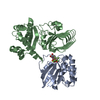


 Mass: 24.305 Da / Num. of mol.: 1 / Source method: obtained synthetically / Formula: Mg
 Mass: 102.975 Da / Num. of mol.: 1 / Source method: obtained synthetically / Formula: AlF4
 Type: RNA linking / Mass: 443.201 Da / Num. of mol.: 1 / Source method: obtained synthetically / Formula: C10H15N5O11P2 / Comment: GDP, energy-carrying molecule*YM
 Sample preparation
 / Beamline: X10SA / Wavelength: 0.9763 Å
 Processing