 Movie
Movie Controller


+ Open data

- Basic information
| Entry | Database: PDB / ID: 3bh6 | ||||||
|---|---|---|---|---|---|---|---|
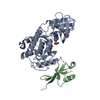
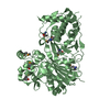
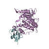
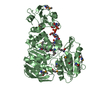
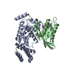
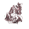
| Title | Crystal structure of the RP2-Arl3 complex bound to GppNHp | ||||||
 Components Components |
| ||||||
 Keywords Keywords | SIGNALING PROTEIN / PROTEIN-PROTEIN COMPLEX / GTPASE ACTIVATING PROTEIN AND GTPASE / RETINITIS PIGMENTOSA / GTP-BINDING / LIPOPROTEIN / MYRISTATE / NUCLEOTIDE-BINDING / DISEASE MUTATION / MEMBRANE / PALMITATE / PHOSPHOPROTEIN / SENSORY TRANSDUCTION / VISION / METAL BINDING PROTEIN | ||||||
| Function / homology |  Function and homology information Function and homology informationTrafficking of myristoylated proteins to the cilium / Trafficking of myristoylated proteins to the cilium / photoreceptor cell development / periciliary membrane compartment / protein localization to ciliary membrane / intraciliary transport / post-Golgi vesicle-mediated transport / photoreceptor connecting cilium / protein localization to cilium / Golgi to plasma membrane transport ...Trafficking of myristoylated proteins to the cilium / Trafficking of myristoylated proteins to the cilium / photoreceptor cell development / periciliary membrane compartment / protein localization to ciliary membrane / intraciliary transport / post-Golgi vesicle-mediated transport / photoreceptor connecting cilium / protein localization to cilium / Golgi to plasma membrane transport / smoothened signaling pathway / small GTPase-mediated signal transduction / ciliary transition zone / mitotic cytokinesis / cilium assembly / axoneme / cytoplasmic microtubule / visual perception / centriole / kidney development / GTPase activator activity / spindle microtubule / : / GDP binding / microtubule cytoskeleton / protein transport / protein folding / cytoplasmic vesicle / midbody / microtubule binding / cilium / ciliary basal body / Golgi membrane / GTPase activity / centrosome / GTP binding / Golgi apparatus / magnesium ion binding / extracellular exosome / nucleoplasm / nucleus / plasma membrane / cytoplasm Similarity search - Function | ||||||
| Biological species |   Homo sapiens (human) Homo sapiens (human) | ||||||
| Method |  X-RAY DIFFRACTION / SYNCHROTRON / MOLECULAR REPLACEMENT / Resolution: 2.6 Å X-RAY DIFFRACTION / SYNCHROTRON / MOLECULAR REPLACEMENT / Resolution: 2.6 Å | ||||||
 Authors Authors | Veltel, S. / Gasper, R. / Wittinghofer, A. | ||||||
 Citation Citation | Journal: Nat.Struct.Mol.Biol. / Year: 2008 Title: The retinitis pigmentosa 2 gene product is a GTPase-activating protein for Arf-like 3 Authors: Veltel, S. / Gasper, R. / Eisenacher, E. / Wittinghofer, A. | ||||||
| History |
|
- Structure visualization
| Structure viewer | Molecule: MolmilJmol/JSmol |
|---|
- Downloads & links
-Download
| PDBx/mmCIF format | 3bh6.cif.gz | 110 KB | Display | PDBx/mmCIF format |
|---|---|---|---|---|
| PDB format | pdb3bh6.ent.gz | 81.7 KB | Display | PDB format |
| PDBx/mmJSON format | 3bh6.json.gz | Tree view | PDBx/mmJSON format | |
| Others |  Other downloads Other downloads |
-Validation report
| Arichive directory | https://data.pdbj.org/pub/pdb/validation_reports/bh/3bh6ftp://data.pdbj.org/pub/pdb/validation_reports/bh/3bh6 | HTTPS FTP |
|---|
-Related structure data
| Related structure data |  3bh7C 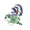 1ksgS 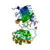 2bx6S S: Starting model for refinement C: citing same article ( |
|---|---|
| Similar structure data |


-Links
 PDBj
PDBj


- Assembly
| Deposited unit | 
| ||||||||
|---|---|---|---|---|---|---|---|---|---|
| 1 |
| ||||||||
| Unit cell |
|
-Components
| #1: Protein | Mass: 18369.828 Da / Num. of mol.: 1 / Fragment: residues 14-177 / Mutation: Q71L Source method: isolated from a genetically manipulated source Source: (gene. exp.)  |
|---|---|
| #2: Protein | Mass: 39827.098 Da / Num. of mol.: 1 Source method: isolated from a genetically manipulated source Source: (gene. exp.) Homo sapiens (human) / Gene: RP2 / Plasmid: pGEX4T3 / Production host: |
| #3: Chemical | ChemComp-MG /   Mass: 24.305 Da / Num. of mol.: 1 / Source method: obtained synthetically / Formula: Mg Mass: 24.305 Da / Num. of mol.: 1 / Source method: obtained synthetically / Formula: Mg |
| #4: Chemical | ChemComp-GNP /   Mass: 522.196 Da / Num. of mol.: 1 / Source method: obtained synthetically / Formula: C10H17N6O13P3 Mass: 522.196 Da / Num. of mol.: 1 / Source method: obtained synthetically / Formula: C10H17N6O13P3Comment: GppNHp, GMPPNP, energy-carrying molecule analogue*YM |
| #5: Water | ChemComp-HOH /  Mass: 18.015 Da / Num. of mol.: 48 / Source method: isolated from a natural source / Formula: H2O Mass: 18.015 Da / Num. of mol.: 48 / Source method: isolated from a natural source / Formula: H2O |
-Experimental details
-Experiment
| Experiment | Method: X-RAY DIFFRACTION / Number of used crystals: 1 |
|---|
- Sample preparation
| Crystal | Density Matthews: 2.45 Å3/Da / Density % sol: 49.8 % |
|---|---|
| Crystal grow | Temperature: 293 K / Method: vapor diffusion, sitting drop / pH: 7.5 Details: 20% PEG 3350, 0.2 M potassium chloride, pH 7.5, VAPOR DIFFUSION, SITTING DROP, temperature 293K |
-Data collection
| Diffraction | Mean temperature: 100 K |
|---|---|
| Diffraction source | Source: SYNCHROTRON / Site: SLS  / Beamline: X10SA / Wavelength: 0.9792 Å / Beamline: X10SA / Wavelength: 0.9792 Å |
| Detector | Type: MARMOSAIC 225 mm CCD / Detector: CCD / Date: Nov 26, 2005 |
| Radiation | Protocol: SINGLE WAVELENGTH / Monochromatic (M) / Laue (L): M / Scattering type: x-ray |
| Radiation wavelength | Wavelength: 0.9792 Å / Relative weight: 1 |
| Reflection | Resolution: 2.6→19.81 Å / Num. all: 18118 / Num. obs: 18118 / % possible obs: 99.6 % / Observed criterion σ(I): 3.8 / Redundancy: 5.9 % / Biso Wilson estimate: 36.068 Å2 / Rsym value: 0.082 / Net I/σ(I): 16.12 |
| Reflection shell | Resolution: 2.6→2.7 Å / Redundancy: 4.6 % / Mean I/σ(I) obs: 3.75 / Num. unique all: 1919 / Rsym value: 0.435 / % possible all: 99.9 |
- Processing
| Software |
| |||||||||||||||||||||||||||||||||||||||||||||||||||||||||||||||||||||||||||||||||||||||||||||||
|---|---|---|---|---|---|---|---|---|---|---|---|---|---|---|---|---|---|---|---|---|---|---|---|---|---|---|---|---|---|---|---|---|---|---|---|---|---|---|---|---|---|---|---|---|---|---|---|---|---|---|---|---|---|---|---|---|---|---|---|---|---|---|---|---|---|---|---|---|---|---|---|---|---|---|---|---|---|---|---|---|---|---|---|---|---|---|---|---|---|---|---|---|---|---|---|---|
| Refinement | Method to determine structure: MOLECULAR REPLACEMENT Starting model: 1KSG, 2BX6 Resolution: 2.6→19.81 Å / Cor.coef. Fo:Fc: 0.924 / Cor.coef. Fo:Fc free: 0.879 / SU B: 11.431 / SU ML: 0.246 / Cross valid method: THROUGHOUT / ESU R: 1.133 / ESU R Free: 0.353 / Stereochemistry target values: MAXIMUM LIKELIHOOD / Details: HYDROGENS HAVE BEEN ADDED IN THE RIDING POSITIONS
| |||||||||||||||||||||||||||||||||||||||||||||||||||||||||||||||||||||||||||||||||||||||||||||||
| Solvent computation | Ion probe radii: 0.8 Å / Shrinkage radii: 0.8 Å / VDW probe radii: 1.2 Å / Solvent model: MASK | |||||||||||||||||||||||||||||||||||||||||||||||||||||||||||||||||||||||||||||||||||||||||||||||
| Displacement parameters | Biso mean: 36.051 Å2
| |||||||||||||||||||||||||||||||||||||||||||||||||||||||||||||||||||||||||||||||||||||||||||||||
| Refinement step | Cycle: LAST / Resolution: 2.6→19.81 Å
| |||||||||||||||||||||||||||||||||||||||||||||||||||||||||||||||||||||||||||||||||||||||||||||||
| Refine LS restraints |
| |||||||||||||||||||||||||||||||||||||||||||||||||||||||||||||||||||||||||||||||||||||||||||||||
| LS refinement shell | Resolution: 2.6→2.666 Å / Total num. of bins used: 20
|
