 Movie
Movie Controller

[English] 日本語
 Yorodumi
- PDB-4d61: Cryo-EM structures of ribosomal 80S complexes with termination fa... -
+ Open data

- Basic information
| Entry | Database: PDB / ID: 4d61 | |||||||||
|---|---|---|---|---|---|---|---|---|---|---|
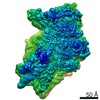
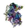
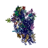
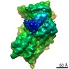
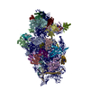
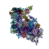
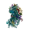
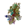
| Title | Cryo-EM structures of ribosomal 80S complexes with termination factors and cricket paralysis virus IRES reveal the IRES in the translocated state | |||||||||
 Components Components |
| |||||||||
 Keywords Keywords | RIBOSOME / CRPV IRES / TERMINATION / RELEASE FACTORS | |||||||||
| Function / homology |  Function and homology information Function and homology informationtranslation termination factor activity / translation release factor complex / cytoplasmic translational termination / regulation of translational termination / translation release factor activity / translation release factor activity, codon specific / protein methylation / sequence-specific mRNA binding / peptidyl-tRNA hydrolase activity / nuclear-transcribed mRNA catabolic process, nonsense-mediated decay ...translation termination factor activity / translation release factor complex / cytoplasmic translational termination / regulation of translational termination / translation release factor activity / translation release factor activity, codon specific / protein methylation / sequence-specific mRNA binding / peptidyl-tRNA hydrolase activity / nuclear-transcribed mRNA catabolic process, nonsense-mediated decay / laminin receptor activity / Protein hydroxylation / Eukaryotic Translation Termination / ubiquitin ligase inhibitor activity / 90S preribosome / Nonsense Mediated Decay (NMD) independent of the Exon Junction Complex (EJC) / positive regulation of signal transduction by p53 class mediator / translational termination / phagocytic cup / Nonsense Mediated Decay (NMD) enhanced by the Exon Junction Complex (EJC) / translation regulator activity / rough endoplasmic reticulum / laminin binding / ribosomal small subunit export from nucleus / gastrulation / MDM2/MDM4 family protein binding / class I DNA-(apurinic or apyrimidinic site) endonuclease activity / cytosolic ribosome / DNA-(apurinic or apyrimidinic site) lyase / maturation of SSU-rRNA from tricistronic rRNA transcript (SSU-rRNA, 5.8S rRNA, LSU-rRNA) / positive regulation of apoptotic signaling pathway / maturation of SSU-rRNA / G1/S transition of mitotic cell cycle / small-subunit processome / spindle / Regulation of expression of SLITs and ROBOs / rRNA processing / rhythmic process / positive regulation of canonical Wnt signaling pathway / regulation of translation / ribosomal small subunit assembly / virus receptor activity / ribosome binding / ribosomal small subunit biogenesis / small ribosomal subunit / small ribosomal subunit rRNA binding / cytosolic small ribosomal subunit / Hydrolases; Acting on acid anhydrides; Acting on GTP to facilitate cellular and subcellular movement / perikaryon / cell differentiation / cytoplasmic translation / mitochondrial inner membrane / postsynaptic density / rRNA binding / structural constituent of ribosome / translation / ribonucleoprotein complex / cell division / DNA repair / mRNA binding / GTPase activity / apoptotic process / centrosome / synapse / dendrite / GTP binding / nucleolus / perinuclear region of cytoplasm / Golgi apparatus / DNA binding / RNA binding / zinc ion binding / membrane / nucleus / plasma membrane / cytoplasm / cytosol Similarity search - Function | |||||||||
| Biological species |  HOMO SAPIENS (human) HOMO SAPIENS (human)  CRICKET PARALYSIS VIRUS CRICKET PARALYSIS VIRUS | |||||||||
| Method | ELECTRON MICROSCOPY / single particle reconstruction / cryo EM / Resolution: 9 Å | |||||||||
 Authors Authors | Muhs, M. / Hilal, T. / Mielke, T. / Skabkin, M.A. / Sanbonmatsu, K.Y. / Pestova, T.V. / Spahn, C.M.T. | |||||||||
 Citation Citation | Journal: Mol Cell / Year: 2015 Title: Cryo-EM of ribosomal 80S complexes with termination factors reveals the translocated cricket paralysis virus IRES. Authors: Margarita Muhs / Tarek Hilal / Thorsten Mielke / Maxim A Skabkin / Karissa Y Sanbonmatsu / Tatyana V Pestova / Christian M T Spahn /   Abstract: The cricket paralysis virus (CrPV) uses an internal ribosomal entry site (IRES) to hijack the ribosome. In a remarkable RNA-based mechanism involving neither initiation factor nor initiator tRNA, the ...The cricket paralysis virus (CrPV) uses an internal ribosomal entry site (IRES) to hijack the ribosome. In a remarkable RNA-based mechanism involving neither initiation factor nor initiator tRNA, the CrPV IRES jumpstarts translation in the elongation phase from the ribosomal A site. Here, we present cryoelectron microscopy (cryo-EM) maps of 80S⋅CrPV-STOP ⋅ eRF1 ⋅ eRF3 ⋅ GMPPNP and 80S⋅CrPV-STOP ⋅ eRF1 complexes, revealing a previously unseen binding state of the IRES and directly rationalizing that an eEF2-dependent translocation of the IRES is required to allow the first A-site occupation. During this unusual translocation event, the IRES undergoes a pronounced conformational change to a more stretched conformation. At the same time, our structural analysis provides information about the binding modes of eRF1 ⋅ eRF3 ⋅ GMPPNP and eRF1 in a minimal system. It shows that neither eRF3 nor ABCE1 are required for the active conformation of eRF1 at the intersection between eukaryotic termination and recycling. | |||||||||
| History |
| |||||||||
| Remark 700 | SHEET DETERMINATION METHOD: DSSP THE SHEETS PRESENTED AS "BB" IN EACH CHAIN ON SHEET RECORDS BELOW ... SHEET DETERMINATION METHOD: DSSP THE SHEETS PRESENTED AS "BB" IN EACH CHAIN ON SHEET RECORDS BELOW IS ACTUALLY AN -7-STRANDED BARREL THIS IS REPRESENTED BY A -6-STRANDED SHEET IN WHICH THE FIRST AND LAST STRANDS ARE IDENTICAL. THE SHEETS PRESENTED AS "BD" IN EACH CHAIN ON SHEET RECORDS BELOW IS ACTUALLY AN -2-STRANDED BARREL THIS IS REPRESENTED BY A -1-STRANDED SHEET IN WHICH THE FIRST AND LAST STRANDS ARE IDENTICAL. |
- Structure visualization
| Movie |
Movie viewer |
|---|---|
| Structure viewer | Molecule: MolmilJmol/JSmol |
- Downloads & links
-Download
| PDBx/mmCIF format | 4d61.cif.gz | 1.9 MB | Display | PDBx/mmCIF format |
|---|---|---|---|---|
| PDB format | pdb4d61.ent.gz | 1.4 MB | Display | PDB format |
| PDBx/mmJSON format | 4d61.json.gz | Tree view | PDBx/mmJSON format | |
| Others |  Other downloads Other downloads |
-Validation report
| Arichive directory | https://data.pdbj.org/pub/pdb/validation_reports/d6/4d61ftp://data.pdbj.org/pub/pdb/validation_reports/d6/4d61 | HTTPS FTP |
|---|
-Related structure data
| Related structure data | 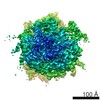 2813MC 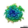 2810C  4d5lC  4d5nC 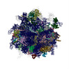 4d5yC 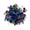 4d67C M: map data used to model this data C: citing same article ( |
|---|---|
| Similar structure data |


-Links
 PDBj
PDBj


- Assembly
| Deposited unit | 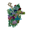
|
|---|---|
| 1 |
|
-Components
-RNA chain , 2 types, 2 molecules 1j
| #1: RNA chain | Mass: 602776.875 Da / Num. of mol.: 1 / Source method: isolated from a natural source / Source: (natural) |
|---|---|
| #37: RNA chain | Mass: 64363.910 Da / Num. of mol.: 1 / Source method: obtained synthetically / Source: (synth.) CRICKET PARALYSIS VIRUS / References: GenBank: AF218039 |
+40S RIBOSOMAL PROTEIN ... , 31 types, 31 molecules ABCDEFGHIJKLMNOPQRSTUVWXYZabcde
-Protein , 2 types, 2 molecules fg
| #33: Protein | Mass: 18004.041 Da / Num. of mol.: 1 / Source method: isolated from a natural source / Source: (natural) |
|---|---|
| #34: Protein | Mass: 35115.652 Da / Num. of mol.: 1 / Source method: isolated from a natural source / Source: (natural) |
-EUKARYOTIC PEPTIDE CHAIN RELEASE FACTOR ... , 2 types, 2 molecules hi
| #35: Protein | Mass: 49040.711 Da / Num. of mol.: 1 Source method: isolated from a genetically manipulated source Source: (gene. exp.) HOMO SAPIENS (human) / Production host:  |
|---|---|
| #36: Protein | Mass: 47888.246 Da / Num. of mol.: 1 / Fragment: G-DOMAIN, UNP RESIDUES 210-635 Source method: isolated from a genetically manipulated source Source: (gene. exp.) HOMO SAPIENS (human) / Production host: |
-Details
| Has protein modification | Y |
|---|---|
| Sequence details | AUTHORS HAVE USED HUMAN SEQUENCE FOR MODEL BUILDING, WITH FOLLOWING PDB-GENBANK OR UNIPROT RESIDUE ...AUTHORS HAVE USED HUMAN SEQUENCE FOR MODEL BUILDING, WITH FOLLOWING PDB-GENBANK OR UNIPROT RESIDUE MAPPING: CHAIN: 1 1-1742 GB X03205.1 1 1742 CHAIN: A 1-295 UNP P08865 1 295 CHAIN: B 1- 264 UNP P61247 1 264 CHAIN: C 1-293 UNP P15880 1 293 CHAIN: D 1-243 UNP P23396 1 243 CHAIN: E 1-263 UNP P22090 1 263 CHAIN: F 1-204 UNP P46782 1 204 CHAIN: G 1-249 UNP P62753 1 249 CHAIN: H 1-194 UNP P62081 1 194 CHAIN: I 1-208 UNP P62241 1 208 CHAIN: J 1-194 UNP P46781 1 194 CHAIN: K 1-165 UNP P46783 1 165 CHAIN: L 1-158 UNP P62280 1 158 CHAIN: M 1-132 UNP P25398 1 132 CHAIN: N 1-151 UNP P62277 1 151 CHAIN: O 1-151 UNP P62263 1 151 CHAIN: P 1-145 UNP P62841 1 145 CHAIN: Q 1-146 UNP P62249 1 146 CHAIN: R 1-135 UNP P08708 1 135 CHAIN: S 1-152 UNP P62269 1 152 CHAIN: T 1-145 UNP P39019 1 145 CHAIN: U 1-119 UNP P60866 1 119 CHAIN: V 1- 83 UNP P63220 1 83 CHAIN: W 1-130 UNP P62244 1 130 CHAIN: X 1-143 UNP P62266 1 143 CHAIN: Y 1-133 UNP P62847 1 133 CHAIN: Z 1-125 UNP P62851 1 125 CHAIN: a 1-115 UNP P62854 1 115 CHAIN: b 1-84 UNP P42677 1 84 CHAIN: c 1-69 UNP P62857 1 69 CHAIN: d 1-56 UNP P62273 1 56 CHAIN: e 1-59 UNP P62861 1 59 CHAIN: f 1-156 UNP P62979 1 156 CHAIN: g 1-317 UNP P63244 1 317 |
-Experimental details
-Experiment
| Experiment | Method: ELECTRON MICROSCOPY |
|---|---|
| EM experiment | Aggregation state: PARTICLE / 3D reconstruction method: single particle reconstruction |
- Sample preparation
| Component | Name: CRICKET PARALYSIS VIRUS IRES RNA BOUND TO MAMMALIAN 80S RIBOSOME, ERF1 AND ERF3 Type: RIBOSOME Details: MICROGRAPHS WERE SELECTED FOR DRIFT AND ASTIGMATISM |
|---|---|
| Buffer solution | Name: 20 MM TRIS PH 7.5, 100 MM KCL, 1 MM DTT, 8.5 MM MGCL2, 0.133 MM GTP, 2.33 MM GMPPNP pH: 7.5 Details: 20 MM TRIS PH 7.5, 100 MM KCL, 1 MM DTT, 8.5 MM MGCL2, 0.133 MM GTP, 2.33 MM GMPPNP |
| Specimen | Conc.: 1.38 mg/ml / Embedding applied: NO / Shadowing applied: NO / Staining applied: NO / Vitrification applied: YES |
| Specimen support | Details: HOLEY CARBON |
| Vitrification | Instrument: FEI VITROBOT MARK II / Cryogen name: ETHANE / Details: LIQUID ETHANE |
- Electron microscopy imaging
| Experimental equipment |  Model: Tecnai F20 / Image courtesy: FEI Company |
|---|---|
| Microscopy | Model: FEI TECNAI F20 / Date: Nov 5, 2012 Details: MICROGRAPHS WERE SELECTED FOR DRIFT AND ASTIGMATISM |
| Electron gun | Electron source:  FIELD EMISSION GUN / Accelerating voltage: 300 kV / Illumination mode: FLOOD BEAM FIELD EMISSION GUN / Accelerating voltage: 300 kV / Illumination mode: FLOOD BEAM |
| Electron lens | Mode: BRIGHT FIELD / Nominal magnification: 115000 X / Calibrated magnification: 194805 X / Nominal defocus max: 5000 nm / Nominal defocus min: 2000 nm / Cs: 2 mm |
| Specimen holder | Temperature: 77 K |
| Image recording | Electron dose: 20 e/Å2 / Film or detector model: TVIPS TEMCAM-F416 (4k x 4k) |
| Image scans | Num. digital images: 1 |
| Radiation wavelength | Relative weight: 1 |
- Processing
| EM software |
| ||||||||||||
|---|---|---|---|---|---|---|---|---|---|---|---|---|---|
| CTF correction | Details: DEFOCUS GROUPS | ||||||||||||
| Symmetry | Point symmetry: C1 (asymmetric) | ||||||||||||
| 3D reconstruction | Method: MULTI-REFERENCE TEMPLATE MATCHING / Resolution: 9 Å / Num. of particles: 64902 / Nominal pixel size: 1.56 Å / Actual pixel size: 1.56 Å Magnification calibration: CROSS- -CORRELATION DENSITIES WITH REFERENCE STRUCTURE Symmetry type: POINT | ||||||||||||
| Atomic model building | Protocol: FLEXIBLE FIT / Space: REAL / Details: METHOD--RIGID BODY, FLEXIBLE FIT | ||||||||||||
| Refinement | Highest resolution: 9 Å | ||||||||||||
| Refinement step | Cycle: LAST / Highest resolution: 9 Å
|
