 Movie
Movie Controller

[English] 日本語
 Yorodumi
- PDB-4d5n: Cryo-EM structures of ribosomal 80S complexes with termination fa... -
+ Open data

- Basic information
| Entry | Database: PDB / ID: 4d5n | |||||||||
|---|---|---|---|---|---|---|---|---|---|---|
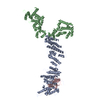
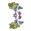
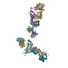
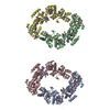
| Title | Cryo-EM structures of ribosomal 80S complexes with termination factors and cricket paralysis virus IRES reveal the IRES in the translocated state | |||||||||
 Components Components |
| |||||||||
 Keywords Keywords | RIBOSOME/RNA / RIBOSOME-RNA COMPLEX / CRPV IRES / RIBOSOME / TERMINATION / RELEASE FACTORS | |||||||||
| Function / homology |  Function and homology information Function and homology informationtranslation termination factor activity / translation release factor complex / cytoplasmic translational termination / regulation of translational termination / translation release factor activity / translation release factor activity, codon specific / protein methylation / sequence-specific mRNA binding / peptidyl-tRNA hydrolase activity / nuclear-transcribed mRNA catabolic process, nonsense-mediated decay ...translation termination factor activity / translation release factor complex / cytoplasmic translational termination / regulation of translational termination / translation release factor activity / translation release factor activity, codon specific / protein methylation / sequence-specific mRNA binding / peptidyl-tRNA hydrolase activity / nuclear-transcribed mRNA catabolic process, nonsense-mediated decay / Protein hydroxylation / Eukaryotic Translation Termination / Nonsense Mediated Decay (NMD) independent of the Exon Junction Complex (EJC) / translational termination / Nonsense Mediated Decay (NMD) enhanced by the Exon Junction Complex (EJC) / cytosolic ribosome / Regulation of expression of SLITs and ROBOs / ribosome binding / RNA binding / cytosol / cytoplasm Similarity search - Function | |||||||||
| Biological species |  HOMO SAPIENS (human) HOMO SAPIENS (human) CRICKET PARALYSIS VIRUS CRICKET PARALYSIS VIRUS | |||||||||
| Method | ELECTRON MICROSCOPY / single particle reconstruction / cryo EM / Resolution: 9 Å | |||||||||
 Authors Authors | Muhs, M. / Hilal, T. / Mielke, T. / Skabkin, M.A. / Sanbonmatsu, K.Y. / Pestova, T.V. / Spahn, C.M.T. | |||||||||
 Citation Citation | Journal: Mol Cell / Year: 2015 Title: Cryo-EM of ribosomal 80S complexes with termination factors reveals the translocated cricket paralysis virus IRES. Authors: Margarita Muhs / Tarek Hilal / Thorsten Mielke / Maxim A Skabkin / Karissa Y Sanbonmatsu / Tatyana V Pestova / Christian M T Spahn /   Abstract: The cricket paralysis virus (CrPV) uses an internal ribosomal entry site (IRES) to hijack the ribosome. In a remarkable RNA-based mechanism involving neither initiation factor nor initiator tRNA, the ...The cricket paralysis virus (CrPV) uses an internal ribosomal entry site (IRES) to hijack the ribosome. In a remarkable RNA-based mechanism involving neither initiation factor nor initiator tRNA, the CrPV IRES jumpstarts translation in the elongation phase from the ribosomal A site. Here, we present cryoelectron microscopy (cryo-EM) maps of 80S⋅CrPV-STOP ⋅ eRF1 ⋅ eRF3 ⋅ GMPPNP and 80S⋅CrPV-STOP ⋅ eRF1 complexes, revealing a previously unseen binding state of the IRES and directly rationalizing that an eEF2-dependent translocation of the IRES is required to allow the first A-site occupation. During this unusual translocation event, the IRES undergoes a pronounced conformational change to a more stretched conformation. At the same time, our structural analysis provides information about the binding modes of eRF1 ⋅ eRF3 ⋅ GMPPNP and eRF1 in a minimal system. It shows that neither eRF3 nor ABCE1 are required for the active conformation of eRF1 at the intersection between eukaryotic termination and recycling. | |||||||||
| History |
|
- Structure visualization
| Movie |
Movie viewer |
|---|---|
| Structure viewer | Molecule: MolmilJmol/JSmol |
- Downloads & links
-Download
| PDBx/mmCIF format | 4d5n.cif.gz | 230 KB | Display | PDBx/mmCIF format |
|---|---|---|---|---|
| PDB format | pdb4d5n.ent.gz | 169.8 KB | Display | PDB format |
| PDBx/mmJSON format | 4d5n.json.gz | Tree view | PDBx/mmJSON format | |
| Others |  Other downloads Other downloads |
-Validation report
| Arichive directory | https://data.pdbj.org/pub/pdb/validation_reports/d5/4d5nftp://data.pdbj.org/pub/pdb/validation_reports/d5/4d5n | HTTPS FTP |
|---|
-Related structure data
| Related structure data | 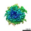 2810MC 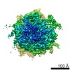 2813C  4d5lC 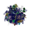 4d5yC  4d61C 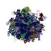 4d67C  4d66 4d68 C: citing same article ( M: map data used to model this data |
|---|---|
| Similar structure data |

-Links
 PDBj
PDBj


- Assembly
| Deposited unit | 
|
|---|---|
| 1 |
|
-Components
| #1: Protein | Mass: 49040.711 Da / Num. of mol.: 1 / Fragment: RESIDUES 5-437 Source method: isolated from a genetically manipulated source Source: (gene. exp.) HOMO SAPIENS (human) / Production host:  |
|---|---|
| #2: RNA chain | Mass: 64363.910 Da / Num. of mol.: 1 / Source method: obtained synthetically / Source: (synth.) CRICKET PARALYSIS VIRUS / References: GenBank: 8895506 |
| Sequence details | FIRST CODING TRIPLET MUTATED TO STOP |
-Experimental details
-Experiment
| Experiment | Method: ELECTRON MICROSCOPY |
|---|---|
| EM experiment | Aggregation state: PARTICLE / 3D reconstruction method: single particle reconstruction |
- Sample preparation
| Component | Name: CRICKET PARALYSIS VIRUS IRES RNA BOUND TO MAMMALIAN 80S RIBOSOME AND ERF1 Type: RIBOSOME / Details: MICROGRAPHS SELECTED FOR ASTIGMATISM AND DRIFT |
|---|---|
| Buffer solution | Name: 20 MM TRIS PH 7.5, 100 MM KCL, 1 MM DTT, 2.5 MM MGCL2, 0.5 MM GTP pH: 7.5 Details: 20 MM TRIS PH 7.5, 100 MM KCL, 1 MM DTT, 2.5 MM MGCL2, 0.5 MM GTP |
| Specimen | Conc.: 1.38 mg/ml / Embedding applied: NO / Shadowing applied: NO / Staining applied: NO / Vitrification applied: YES |
| Specimen support | Details: HOLEY CARBON |
| Vitrification | Instrument: FEI VITROBOT MARK II / Cryogen name: ETHANE / Details: LIQUID ETHANE |
- Electron microscopy imaging
| Experimental equipment |  Model: Tecnai F20 / Image courtesy: FEI Company |
|---|---|
| Microscopy | Model: FEI TECNAI F20 / Date: Apr 17, 2012 Details: GOOD MICROGRAPHS SELECTED FOR ASTIGMATISM AND DRIFT |
| Electron gun | Electron source:  FIELD EMISSION GUN / Accelerating voltage: 300 kV / Illumination mode: FLOOD BEAM FIELD EMISSION GUN / Accelerating voltage: 300 kV / Illumination mode: FLOOD BEAM |
| Electron lens | Mode: BRIGHT FIELD / Nominal magnification: 39000 X / Calibrated magnification: 65520 X / Nominal defocus max: 4000 nm / Nominal defocus min: 2000 nm / Cs: 2 mm |
| Specimen holder | Temperature: 77 K |
| Image recording | Electron dose: 20 e/Å2 / Film or detector model: KODAK SO-163 FILM |
| Image scans | Num. digital images: 366 |
- Processing
| EM software |
| ||||||||||||
|---|---|---|---|---|---|---|---|---|---|---|---|---|---|
| CTF correction | Details: DEFOCUS GROUPS | ||||||||||||
| Symmetry | Point symmetry: C1 (asymmetric) | ||||||||||||
| 3D reconstruction | Method: MULTI-REFERENCE TEMPLATE MATCHING / Resolution: 9 Å / Num. of particles: 109596 / Nominal pixel size: 1.56 Å / Actual pixel size: 1.56 Å Magnification calibration: CROSS- -CORRELATION DENSITIES WITH REFERENCE STRUCTURE Details: SUBMISSION BASED ON EXPERIMENTAL DATA FROM EMDB EMD-2810. (DEPOSITION ID: 12907). Symmetry type: POINT | ||||||||||||
| Atomic model building | Protocol: FLEXIBLE FIT / Space: REAL / Details: METHOD--RIGID BODY, FLEXIBLE FIT | ||||||||||||
| Refinement | Highest resolution: 9 Å | ||||||||||||
| Refinement step | Cycle: LAST / Highest resolution: 9 Å
|
