 Movie
Movie Controller

 Yorodumi
+ Open data

- Basic information
| Entry | Database: EMDB / ID: EMD-2810 | |||||||||
|---|---|---|---|---|---|---|---|---|---|---|
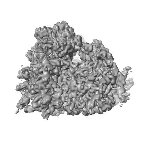
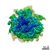
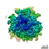
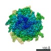
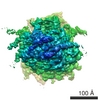
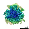
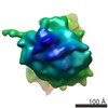
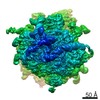
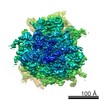
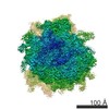
| Title | Cryo-EM structures of ribosomal 80S complexes with termination factors and cricket paralysis virus IRES reveal the IRES in the translocated state | |||||||||
 Map data Map data | Reconstruction of mammalian termination complex with CrPV IRES-RNA and eRF1 | |||||||||
 Sample Sample |
| |||||||||
 Keywords Keywords | CrPV IRES / ribosome / Termination / release factors | |||||||||
| Function / homology |  Function and homology information Function and homology informationtranslation termination factor activity / translation release factor complex / cytoplasmic translational termination / regulation of translational termination / translation release factor activity / translation release factor activity, codon specific / protein methylation / sequence-specific mRNA binding / peptidyl-tRNA hydrolase activity / nuclear-transcribed mRNA catabolic process, nonsense-mediated decay ...translation termination factor activity / translation release factor complex / cytoplasmic translational termination / regulation of translational termination / translation release factor activity / translation release factor activity, codon specific / protein methylation / sequence-specific mRNA binding / peptidyl-tRNA hydrolase activity / nuclear-transcribed mRNA catabolic process, nonsense-mediated decay / laminin receptor activity / Protein hydroxylation / Eukaryotic Translation Termination / ubiquitin ligase inhibitor activity / 90S preribosome / Nonsense Mediated Decay (NMD) independent of the Exon Junction Complex (EJC) / positive regulation of signal transduction by p53 class mediator / translational termination / phagocytic cup / Nonsense Mediated Decay (NMD) enhanced by the Exon Junction Complex (EJC) / translation regulator activity / rough endoplasmic reticulum / ribosomal small subunit export from nucleus / laminin binding / gastrulation / MDM2/MDM4 family protein binding / class I DNA-(apurinic or apyrimidinic site) endonuclease activity / cytosolic ribosome / DNA-(apurinic or apyrimidinic site) lyase / maturation of LSU-rRNA from tricistronic rRNA transcript (SSU-rRNA, 5.8S rRNA, LSU-rRNA) / maturation of SSU-rRNA from tricistronic rRNA transcript (SSU-rRNA, 5.8S rRNA, LSU-rRNA) / positive regulation of apoptotic signaling pathway / maturation of SSU-rRNA / small-subunit processome / spindle / Regulation of expression of SLITs and ROBOs / rRNA processing / rhythmic process / positive regulation of canonical Wnt signaling pathway / regulation of translation / ribosomal small subunit assembly / virus receptor activity / ribosome binding / ribosomal small subunit biogenesis / small ribosomal subunit / small ribosomal subunit rRNA binding / cytosolic small ribosomal subunit / cytosolic large ribosomal subunit / perikaryon / cell differentiation / cytoplasmic translation / mitochondrial inner membrane / postsynaptic density / rRNA binding / structural constituent of ribosome / translation / ribonucleoprotein complex / cell division / DNA repair / mRNA binding / apoptotic process / centrosome / synapse / dendrite / perinuclear region of cytoplasm / nucleolus / Golgi apparatus / DNA binding / RNA binding / zinc ion binding / membrane / nucleus / plasma membrane / cytoplasm / cytosol Similarity search - Function | |||||||||
| Biological species |   Homo sapiens (human) / Homo sapiens (human) /  Cricket paralysis virus Cricket paralysis virus | |||||||||
| Method | single particle reconstruction / cryo EM / Resolution: 8.7 Å | |||||||||
 Authors Authors | Muhs M / Hilal T / Mielke T / Skabkin MA / Sanbonmatsu KY / Pestova TV / Spahn CMT | |||||||||
 Citation Citation | Journal: Mol Cell / Year: 2015 Title: Cryo-EM of ribosomal 80S complexes with termination factors reveals the translocated cricket paralysis virus IRES. Authors: Margarita Muhs / Tarek Hilal / Thorsten Mielke / Maxim A Skabkin / Karissa Y Sanbonmatsu / Tatyana V Pestova / Christian M T Spahn /   Abstract: The cricket paralysis virus (CrPV) uses an internal ribosomal entry site (IRES) to hijack the ribosome. In a remarkable RNA-based mechanism involving neither initiation factor nor initiator tRNA, the ...The cricket paralysis virus (CrPV) uses an internal ribosomal entry site (IRES) to hijack the ribosome. In a remarkable RNA-based mechanism involving neither initiation factor nor initiator tRNA, the CrPV IRES jumpstarts translation in the elongation phase from the ribosomal A site. Here, we present cryoelectron microscopy (cryo-EM) maps of 80S⋅CrPV-STOP ⋅ eRF1 ⋅ eRF3 ⋅ GMPPNP and 80S⋅CrPV-STOP ⋅ eRF1 complexes, revealing a previously unseen binding state of the IRES and directly rationalizing that an eEF2-dependent translocation of the IRES is required to allow the first A-site occupation. During this unusual translocation event, the IRES undergoes a pronounced conformational change to a more stretched conformation. At the same time, our structural analysis provides information about the binding modes of eRF1 ⋅ eRF3 ⋅ GMPPNP and eRF1 in a minimal system. It shows that neither eRF3 nor ABCE1 are required for the active conformation of eRF1 at the intersection between eukaryotic termination and recycling. | |||||||||
| History |
|
- Structure visualization
| Movie |
Movie viewer |
|---|---|
| Structure viewer | EM map: SurfViewMolmilJmol/JSmol |
| Supplemental images |

- Downloads & links
-EMDB archive
| Map data | emd_2810.map.gz | 86.3 MB | EMDB map data format | |
|---|---|---|---|---|
| Header (meta data) | emd-2810-v30.xmlemd-2810.xml | 19.2 KB 19.2 KB | Display Display | EMDB header |
| Images | 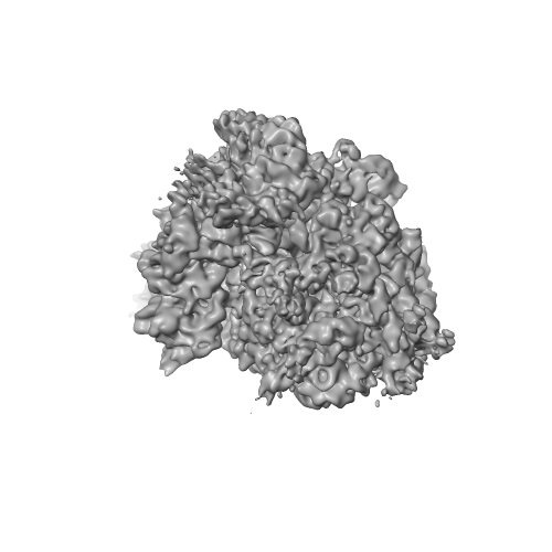 eRF1.png eRF1.png emd_2810.png emd_2810.png | 80.3 KB 119.6 KB | ||
| Archive directory |  http://ftp.pdbj.org/pub/emdb/structures/EMD-2810ftp://ftp.pdbj.org/pub/emdb/structures/EMD-2810 http://ftp.pdbj.org/pub/emdb/structures/EMD-2810ftp://ftp.pdbj.org/pub/emdb/structures/EMD-2810 | HTTPS FTP |
-Related structure data
| Related structure data |  4d5lMC  4d5nMC  4d5yMC  2813C  4d61C  4d67C M: atomic model generated by this map C: citing same article ( |
|---|---|
| Similar structure data |

-Links
| EMDB pages | EMDB (EBI/PDBe) / EMDataResource |
|---|---|
| Related items in Molecule of the Month |


-Map
| File | Download / File: emd_2810.map.gz / Format: CCP4 / Size: 100.6 MB / Type: IMAGE STORED AS FLOATING POINT NUMBER (4 BYTES) | ||||||||||||||||||||||||||||||||||||||||||||||||||||||||||||||||||||
|---|---|---|---|---|---|---|---|---|---|---|---|---|---|---|---|---|---|---|---|---|---|---|---|---|---|---|---|---|---|---|---|---|---|---|---|---|---|---|---|---|---|---|---|---|---|---|---|---|---|---|---|---|---|---|---|---|---|---|---|---|---|---|---|---|---|---|---|---|---|
| Annotation | Reconstruction of mammalian termination complex with CrPV IRES-RNA and eRF1 | ||||||||||||||||||||||||||||||||||||||||||||||||||||||||||||||||||||
| Projections & slices | Image control
Images are generated by Spider. | ||||||||||||||||||||||||||||||||||||||||||||||||||||||||||||||||||||
| Voxel size | X=Y=Z: 1.56 Å | ||||||||||||||||||||||||||||||||||||||||||||||||||||||||||||||||||||
| Density |
| ||||||||||||||||||||||||||||||||||||||||||||||||||||||||||||||||||||
| Symmetry | Space group: 1 | ||||||||||||||||||||||||||||||||||||||||||||||||||||||||||||||||||||
| Details | EMDB XML:
CCP4 map header:
| ||||||||||||||||||||||||||||||||||||||||||||||||||||||||||||||||||||
 Z (Sec.)
 Y (Row.)
 X (Col.)


-Supplemental data
- Sample components
-Entire : Ribosomal 80S termination complex with CrPV IRES-RNA and eRF1
| Entire | Name: Ribosomal 80S termination complex with CrPV IRES-RNA and eRF1 |
|---|---|
| Components |
|
-Supramolecule #1000: Ribosomal 80S termination complex with CrPV IRES-RNA and eRF1
| Supramolecule | Name: Ribosomal 80S termination complex with CrPV IRES-RNA and eRF1 type: sample / ID: 1000 Oligomeric state: CrPV IRES and eRF1 bound to one 80S ribosome Number unique components: 3 |
|---|---|
| Molecular weight | Theoretical: 4.5 MDa |
-Supramolecule #1: 80S ribosome
| Supramolecule | Name: 80S ribosome / type: complex / ID: 1 / Recombinant expression: No / Ribosome-details: ribosome-eukaryote: ALL |
|---|---|
| Source (natural) | Organism: |
| Molecular weight | Theoretical: 4.5 MDa |
-Macromolecule #1: eukaryoric release factor 1
| Macromolecule | Name: eukaryoric release factor 1 / type: protein_or_peptide / ID: 1 / Name.synonym: eRF1 / Number of copies: 1 / Oligomeric state: Monomer / Recombinant expression: Yes |
|---|---|
| Source (natural) | Organism: Homo sapiens (human) / synonym: Human |
| Molecular weight | Theoretical: 50 KDa |
| Recombinant expression | Organism:  |
| Sequence | UniProtKB: Eukaryotic peptide chain release factor subunit 1 |
-Macromolecule #2: Cricket paralysis virus IRES RNA
| Macromolecule | Name: Cricket paralysis virus IRES RNA / type: rna / ID: 2 / Name.synonym: CrPV IRES RNA / Classification: OTHER / Structure: OTHER / Synthetic?: No |
|---|---|
| Source (natural) | Organism: Cricket paralysis virus |
| Sequence | String: AAAAAUGUGA UCUUGCUUGU AAAUACAAUU UUGAGAGGUU AAUAAAUUAC AAGUAGUGCU AUUUUUGUAU UUAGGUUAGC UAUUUAGCUU UACGUUCCAG GAUGCCUAGU GGCAGCCCCA CAAUAUCCAG GAAGCCCUCU CUGCGGUUUU UCAGAUUAGG UAGUCGAAAA ...String: AAAAAUGUGA UCUUGCUUGU AAAUACAAUU UUGAGAGGUU AAUAAAUUAC AAGUAGUGCU AUUUUUGUAU UUAGGUUAGC UAUUUAGCUU UACGUUCCAG GAUGCCUAGU GGCAGCCCCA CAAUAUCCAG GAAGCCCUCU CUGCGGUUUU UCAGAUUAGG UAGUCGAAAA ACCUAAGAAA UUUACCUUAA GGCUUCCUCG A |
-Experimental details
-Structure determination
| Method | cryo EM |
|---|---|
 Processing Processing | single particle reconstruction |
| Aggregation state | particle |
-Sample preparation
| Concentration | 1.38 mg/mL |
|---|---|
| Buffer | pH: 7.5 Details: 20 mM Tris pH 7.5, 100 mM KCl, 1 mM DTT, 2.5 mM MgCl2, 0.5 mM GTP |
| Grid | Details: Quantifoil grids with additional continuous carbon support |
| Vitrification | Cryogen name: ETHANE / Chamber humidity: 85 % / Instrument: FEI VITROBOT MARK II / Method: blot for 2/4 seconds before plunging |
- Electron microscopy
| Microscope | FEI TECNAI F20 |
|---|---|
| Temperature | Min: 77 K |
| Alignment procedure | Legacy - Astigmatism: Objective lens astigmatism was corrected at 200,000 times magnification |
| Details | minimal dose system |
| Date | Apr 17, 2012 |
| Image recording | Category: FILM / Film or detector model: KODAK SO-163 FILM / Digitization - Scanner: OTHER / Number real images: 366 / Average electron dose: 20 e/Å2 / Bits/pixel: 16 |
| Electron beam | Acceleration voltage: 300 kV / Electron source:  FIELD EMISSION GUN FIELD EMISSION GUN |
| Electron optics | Calibrated magnification: 65520 / Illumination mode: FLOOD BEAM / Imaging mode: BRIGHT FIELD / Cs: 2 mm / Nominal defocus max: 4.0 µm / Nominal defocus min: 2.0 µm / Nominal magnification: 39000 |
| Sample stage | Specimen holder model: GATAN LIQUID NITROGEN |
| Experimental equipment |  Model: Tecnai F20 / Image courtesy: FEI Company |
-Image processing
| Details | The particles were selected using SIGNATURE and processed by using SPIDER and SPARX |
|---|---|
| CTF correction | Details: Defocus group |
| Final reconstruction | Applied symmetry - Point group: C1 (asymmetric) / Algorithm: OTHER / Resolution.type: BY AUTHOR / Resolution: 8.7 Å / Resolution method: OTHER / Software - Name: SPIDER, SPARX Details: The particles were selected using SIGNATURE and processed by using SPIDER and SPARX. Number images used: 109596 |
-Atomic model buiding 1
| Initial model | PDB ID:  4cxc Chain - #0 - Chain ID: A / Chain - #1 - Chain ID: B / Chain - #2 - Chain ID: C / Chain - #3 - Chain ID: D / Chain - #4 - Chain ID: E / Chain - #5 - Chain ID: F / Chain - #6 - Chain ID: G / Chain - #7 - Chain ID: H / Chain - #8 - Chain ID: I / Chain - #9 - Chain ID: J / Chain - #10 - Chain ID: K / Chain - #11 - Chain ID: L / Chain - #12 - Chain ID: M / Chain - #13 - Chain ID: N / Chain - #14 - Chain ID: O / Chain - #15 - Chain ID: P / Chain - #16 - Chain ID: Q / Chain - #17 - Chain ID: R / Chain - #18 - Chain ID: S / Chain - #19 - Chain ID: T / Chain - #20 - Chain ID: U / Chain - #21 - Chain ID: V / Chain - #22 - Chain ID: W / Chain - #23 - Chain ID: X / Chain - #24 - Chain ID: Y / Chain - #25 - Chain ID: Z / Chain - #26 - Chain ID: a / Chain - #27 - Chain ID: b / Chain - #28 - Chain ID: c / Chain - #29 - Chain ID: d / Chain - #30 - Chain ID: e / Chain - #31 - Chain ID: f / Chain - #32 - Chain ID: g / Chain - #33 - Chain ID: 1 |
|---|---|
| Software | Name: CHIMERA |
| Refinement | Space: REAL / Protocol: RIGID BODY FIT |
| Output model | PDB-4d5l: PDB-4d5n: PDB-4d5y: |
-Atomic model buiding 2
| Initial model | PDB ID: 4cxd Chain - #0 - Chain ID: A / Chain - #1 - Chain ID: B / Chain - #2 - Chain ID: C / Chain - #3 - Chain ID: D / Chain - #4 - Chain ID: E / Chain - #5 - Chain ID: F / Chain - #6 - Chain ID: G / Chain - #7 - Chain ID: H / Chain - #8 - Chain ID: I / Chain - #9 - Chain ID: J / Chain - #10 - Chain ID: L / Chain - #11 - Chain ID: M / Chain - #12 - Chain ID: N / Chain - #13 - Chain ID: O / Chain - #14 - Chain ID: P / Chain - #15 - Chain ID: Q / Chain - #16 - Chain ID: R / Chain - #17 - Chain ID: S / Chain - #18 - Chain ID: T / Chain - #19 - Chain ID: U / Chain - #20 - Chain ID: V / Chain - #21 - Chain ID: W / Chain - #22 - Chain ID: X / Chain - #23 - Chain ID: Y / Chain - #24 - Chain ID: Z / Chain - #25 - Chain ID: a / Chain - #26 - Chain ID: b / Chain - #27 - Chain ID: c / Chain - #28 - Chain ID: d / Chain - #29 - Chain ID: e / Chain - #30 - Chain ID: f / Chain - #31 - Chain ID: g / Chain - #32 - Chain ID: h / Chain - #33 - Chain ID: i / Chain - #34 - Chain ID: j / Chain - #35 - Chain ID: k / Chain - #36 - Chain ID: l / Chain - #37 - Chain ID: m / Chain - #38 - Chain ID: n / Chain - #39 - Chain ID: o / Chain - #40 - Chain ID: p / Chain - #41 - Chain ID: t / Chain - #42 - Chain ID: u |
|---|---|
| Software | Name: CHIMERA |
| Refinement | Space: REAL / Protocol: RIGID BODY FIT |
| Output model | PDB-4d5l: PDB-4d5n: PDB-4d5y: |
