Movie
Movie Controller


+ Open data

- Basic information
| Entry | Database: EMDB / ID: EMD-1480 | |||||||||
|---|---|---|---|---|---|---|---|---|---|---|
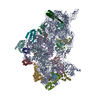
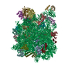
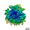
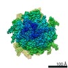
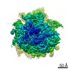
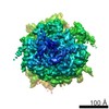
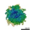
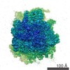
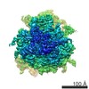
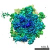
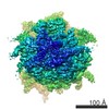
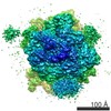
| Title | 3D structure of the canine 80S ribosome | |||||||||
 Map data Map data | 3D volume of the canine 80S ribosome determined at 8.7 A resolution (Fsc 0.5), within the context of a ribosome-channel complex from ER membranes. | |||||||||
 Sample Sample |
| |||||||||
 Keywords Keywords | eukaryotic ribosome / protein translation / tRNA translocation / expansion segments | |||||||||
| Biological species |  | |||||||||
| Method | single particle reconstruction / cryo EM / Resolution: 8.7 Å | |||||||||
 Authors Authors | Chandramouli P / Topf M / Menetret JF / Eswar N / Cannone JJ / Gutell R / Sali A / Akey CW | |||||||||
 Citation Citation | Journal: Structure / Year: 2008 Title: Structure of the mammalian 80S ribosome at 8.7 A resolution. Authors: Preethi Chandramouli / Maya Topf / Jean-François Ménétret / Narayanan Eswar / Jamie J Cannone / Robin R Gutell / Andrej Sali / Christopher W Akey /  Abstract: In this paper, we present a structure of the mammalian ribosome determined at approximately 8.7 A resolution by electron cryomicroscopy and single-particle methods. A model of the ribosome was ...In this paper, we present a structure of the mammalian ribosome determined at approximately 8.7 A resolution by electron cryomicroscopy and single-particle methods. A model of the ribosome was created by docking homology models of subunit rRNAs and conserved proteins into the density map. We then modeled expansion segments in the subunit rRNAs and found unclaimed density for approximately 20 proteins. In general, many conserved proteins and novel proteins interact with expansion segments to form an integrated framework that may stabilize the mature ribosome. Our structure provides a snapshot of the mammalian ribosome at the beginning of translation and lends support to current models in which large movements of the small subunit and L1 stalk occur during tRNA translocation. Finally, details are presented for intersubunit bridges that are specific to the eukaryotic ribosome. We suggest that these bridges may help reset the conformation of the ribosome to prepare for the next cycle of chain elongation. | |||||||||
| History |
|
- Structure visualization
| Movie |
 Movie viewer Movie viewer |
|---|---|
| Structure viewer | EM map: SurfViewMolmilJmol/JSmol |
| Supplemental images |

- Downloads & links
-EMDB archive
| Map data | emd_1480.map.gz | 4 MB | EMDB map data format | |
|---|---|---|---|---|
| Header (meta data) | emd-1480-v30.xmlemd-1480.xml | 10.4 KB 10.4 KB | Display Display | EMDB header |
| Images | 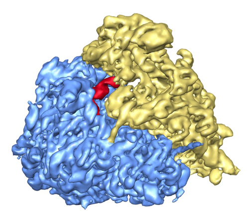 1480.jpg 1480.jpg | 169.3 KB | ||
| Archive directory |  http://ftp.pdbj.org/pub/emdb/structures/EMD-1480ftp://ftp.pdbj.org/pub/emdb/structures/EMD-1480 http://ftp.pdbj.org/pub/emdb/structures/EMD-1480ftp://ftp.pdbj.org/pub/emdb/structures/EMD-1480 | HTTPS FTP |
-Related structure data
| Related structure data | 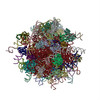 4v5zMC M: atomic model generated by this map C: citing same article ( |
|---|---|
| Similar structure data |
-Links
| EMDB pages | EMDB (EBI/PDBe) / EMDataResource |
|---|---|
| Related items in Molecule of the Month |

-Map
| File | Download / File: emd_1480.map.gz / Format: CCP4 / Size: 17.7 MB / Type: IMAGE STORED AS FLOATING POINT NUMBER (4 BYTES) | ||||||||||||||||||||||||||||||||||||||||||||||||||||||||||||||||||||
|---|---|---|---|---|---|---|---|---|---|---|---|---|---|---|---|---|---|---|---|---|---|---|---|---|---|---|---|---|---|---|---|---|---|---|---|---|---|---|---|---|---|---|---|---|---|---|---|---|---|---|---|---|---|---|---|---|---|---|---|---|---|---|---|---|---|---|---|---|---|
| Annotation | 3D volume of the canine 80S ribosome determined at 8.7 A resolution (Fsc 0.5), within the context of a ribosome-channel complex from ER membranes. | ||||||||||||||||||||||||||||||||||||||||||||||||||||||||||||||||||||
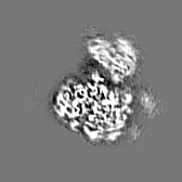
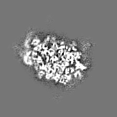
| Projections & slices | Image control
Images are generated by Spider. | ||||||||||||||||||||||||||||||||||||||||||||||||||||||||||||||||||||
| Voxel size | X=Y=Z: 2.73 Å | ||||||||||||||||||||||||||||||||||||||||||||||||||||||||||||||||||||
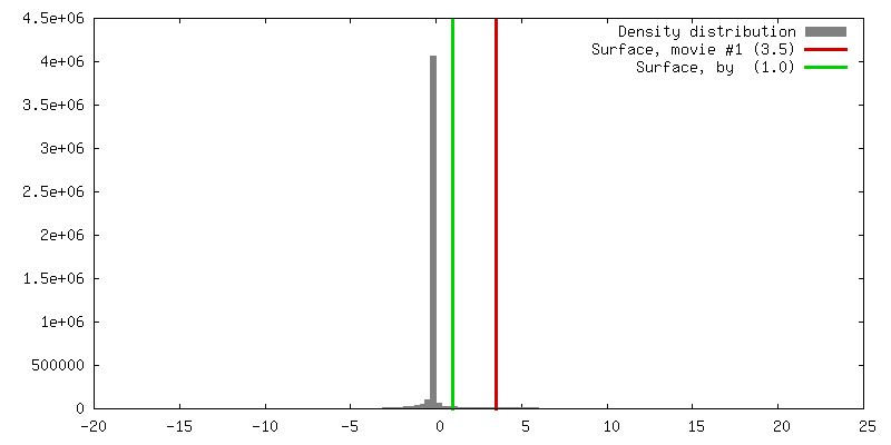
| Density |
| ||||||||||||||||||||||||||||||||||||||||||||||||||||||||||||||||||||
| Symmetry | Space group: 1 | ||||||||||||||||||||||||||||||||||||||||||||||||||||||||||||||||||||
| Details | EMDB XML:
CCP4 map header:
| ||||||||||||||||||||||||||||||||||||||||||||||||||||||||||||||||||||
 Z (Sec.)
 Y (Row.)
 X (Col.)


-Supplemental data
- Sample components
-Entire : canine 80S ribosome
| Entire | Name: canine 80S ribosome |
|---|---|
| Components |
|
-Supramolecule #1000: canine 80S ribosome
| Supramolecule | Name: canine 80S ribosome / type: sample / ID: 1000 Details: Sample was monodisperse with some mild aggregation. Oligomeric state: monomer / Number unique components: 1 |
|---|---|
| Molecular weight | Theoretical: 3.6 MDa |
-Supramolecule #1: 80S ribosome
| Supramolecule | Name: 80S ribosome / type: complex / ID: 1 / Name.synonym: ribosome Details: The ribosome structure was determined within a larger, ribosome-channel complex isolated from ER membranes. Recombinant expression: No / Ribosome-details: ribosome-eukaryote: ALL |
|---|---|
| Source (natural) | Organism: |
| Molecular weight | Theoretical: 3.6 MDa |
-Experimental details
-Structure determination
| Method | cryo EM |
|---|---|
 Processing Processing | single particle reconstruction |
| Aggregation state | particle |
-Sample preparation
| Buffer | pH: 7.5 Details: 30mM Hepesm 50mM KAc, 10mM Mg acetate and 1.5% digitonin. |
|---|---|
| Grid | Details: 400 |
| Vitrification | Cryogen name: ETHANE / Chamber humidity: 90 % / Instrument: HOMEMADE PLUNGER Details: Vitrification instrument: home made plunger. in cold room Method: blot for 1 second |
- Electron microscopy
| Microscope | FEI TECNAI 20 |
|---|---|
| Temperature | Average: 93 K |
| Details | data were collected on Oxford and a Gatan cryo-holders |
| Image recording | Category: FILM / Film or detector model: KODAK SO-163 FILM / Digitization - Scanner: OTHER / Digitization - Sampling interval: 4.5 µm / Number real images: 500 / Average electron dose: 20 e/Å2 / Details: Creoscitex Eversmart was used to scan negatives. / Od range: 1 / Bits/pixel: 16 |
| Electron beam | Acceleration voltage: 200 kV / Electron source:  FIELD EMISSION GUN FIELD EMISSION GUN |
| Electron optics | Calibrated magnification: 51000 / Illumination mode: FLOOD BEAM / Imaging mode: BRIGHT FIELD / Cs: 2 mm / Nominal defocus max: 3.0 µm / Nominal defocus min: 0.5 µm / Nominal magnification: 50000 |
| Sample stage | Specimen holder: eucentric / Specimen holder model: OTHER |
-Image processing
| Details | Particles selected with Boxer |
|---|---|
| CTF correction | Details: by micrograph |
| Final reconstruction | Applied symmetry - Point group: C1 (asymmetric) / Algorithm: OTHER / Resolution.type: BY AUTHOR / Resolution: 8.7 Å / Resolution method: FSC 0.5 CUT-OFF / Software - Name: EMAN Details: sep option equals 3 and setsf were used in the final cycle. a combined structure factor was used to correct for amplitudes in EMAN using the low resolution region from the images and the mid- ...Details: sep option equals 3 and setsf were used in the final cycle. a combined structure factor was used to correct for amplitudes in EMAN using the low resolution region from the images and the mid-resolution region from a low angle X-ray diffraction pattern. Number images used: 78800 |
| Final angle assignment | Details: 2 degree steps between classes |