Movie
Movie Controller

[English] 日本語
 Yorodumi
- PDB-4v5z: Structure of a mammalian 80S ribosome obtained by docking homolog... -
+ Open data

- Basic information
| Entry | Database: PDB / ID: 4v5z | |||||||||
|---|---|---|---|---|---|---|---|---|---|---|
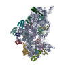
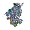
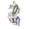
| Title | Structure of a mammalian 80S ribosome obtained by docking homology models of the RNA and proteins into an 8.7 A cryo-EM map | |||||||||
 Components Components |
| |||||||||
 Keywords Keywords | RIBOSOMAL PROTEIN/RNA / protein-RNA complex / 40S ribosomal subunit / RIBOSOMAL PROTEIN-RNA COMPLEX | |||||||||
| Function / homology | RNA / RNA (> 10) / RNA (> 100) / RNA (> 1000) Function and homology information Function and homology information | |||||||||
| Biological species |  | |||||||||
| Method | ELECTRON MICROSCOPY / single particle reconstruction / cryo EM / Resolution: 8.7 Å | |||||||||
 Authors Authors | Chandramouli, P. / Akey, C.W. | |||||||||
 Citation Citation | Journal: Structure / Year: 2008 Title: Structure of the mammalian 80S ribosome at 8.7 A resolution. Authors: Preethi Chandramouli / Maya Topf / Jean-François Ménétret / Narayanan Eswar / Jamie J Cannone / Robin R Gutell / Andrej Sali / Christopher W Akey /  Abstract: In this paper, we present a structure of the mammalian ribosome determined at approximately 8.7 A resolution by electron cryomicroscopy and single-particle methods. A model of the ribosome was ...In this paper, we present a structure of the mammalian ribosome determined at approximately 8.7 A resolution by electron cryomicroscopy and single-particle methods. A model of the ribosome was created by docking homology models of subunit rRNAs and conserved proteins into the density map. We then modeled expansion segments in the subunit rRNAs and found unclaimed density for approximately 20 proteins. In general, many conserved proteins and novel proteins interact with expansion segments to form an integrated framework that may stabilize the mature ribosome. Our structure provides a snapshot of the mammalian ribosome at the beginning of translation and lends support to current models in which large movements of the small subunit and L1 stalk occur during tRNA translocation. Finally, details are presented for intersubunit bridges that are specific to the eukaryotic ribosome. We suggest that these bridges may help reset the conformation of the ribosome to prepare for the next cycle of chain elongation. | |||||||||
| History |
|
- Structure visualization
| Movie |
Movie viewer |
|---|---|
| Structure viewer | Molecule: MolmilJmol/JSmol |
- Downloads & links
-Download
| PDBx/mmCIF format | 4v5z.cif.gz | 1.3 MB | Display | PDBx/mmCIF format |
|---|---|---|---|---|
| PDB format | pdb4v5z.ent.gz | Display | PDB format | |
| PDBx/mmJSON format | 4v5z.json.gz | Tree view | PDBx/mmJSON format | |
| Others |  Other downloads Other downloads |
-Validation report
| Arichive directory | https://data.pdbj.org/pub/pdb/validation_reports/v5/4v5zftp://data.pdbj.org/pub/pdb/validation_reports/v5/4v5z | HTTPS FTP |
|---|
-Related structure data
| Related structure data | 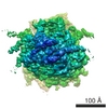 1480MC M: map data used to model this data C: citing same article ( |
|---|---|
| Similar structure data |


-Links
 PDBj
PDBj


- Assembly
| Deposited unit | 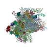
|
|---|---|
| 1 |
|
-Components
-RNA chain , 8 types, 8 molecules AAAGAHB1B0BXBYBZ
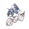
| #1: RNA chain | Mass: 504281.344 Da / Num. of mol.: 1 / Source method: isolated from a natural source Details: Ribosome-channel complexes isolated from ER membranes were used to create a 3D map which was used to model the 40S subunit in the 80S ribosome. Source: (natural) |
|---|---|
| #7: RNA chain | Mass: 4539.804 Da / Num. of mol.: 1 / Source method: isolated from a natural source / Source: (natural) |
| #8: RNA chain | Mass: 13335.024 Da / Num. of mol.: 1 / Source method: isolated from a natural source / Source: (natural) |
| #25: RNA chain | Mass: 39561.457 Da / Num. of mol.: 1 / Source method: isolated from a natural source / Source: (natural) |
| #26: RNA chain | Mass: 940211.062 Da / Num. of mol.: 1 / Source method: isolated from a natural source Details: Ribosome-channel complexes isolated from ER membranes were used to create a 3D map which was used to model the 60S subunit in the 80S ribosome. Source: (natural) |
| #50: RNA chain | Mass: 36425.715 Da / Num. of mol.: 1 / Source method: isolated from a natural source / Source: (natural) |
| #51: RNA chain | Mass: 37084.961 Da / Num. of mol.: 1 / Source method: isolated from a natural source / Source: (natural) |
| #52: RNA chain | Mass: 23175.754 Da / Num. of mol.: 1 / Source method: isolated from a natural source / Source: (natural) |
+RNA Expansion segment ... , 28 types, 28 molecules ABACADAEAFBABBBCBDBEBFBGBHBIBJBKBLBMBNBOBPBQBRBSBTBUBVBW
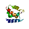
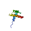
-40S Ribosomal protein ... , 16 types, 16 molecules AaAbAcAdAeAgAhAiAjAkAlAmAnAoAqAs
| #9: Protein | Mass: 35115.652 Da / Num. of mol.: 1 / Source method: isolated from a natural source / Source: (natural) |
|---|---|
| #10: Protein | Mass: 32913.965 Da / Num. of mol.: 1 / Source method: isolated from a natural source / Source: (natural) |
| #11: Protein | Mass: 26729.369 Da / Num. of mol.: 1 / Source method: isolated from a natural source / Source: (natural) |
| #12: Protein | Mass: 24373.447 Da / Num. of mol.: 1 / Source method: isolated from a natural source / Source: (natural) |
| #13: Protein | Mass: 19397.674 Da / Num. of mol.: 1 / Source method: isolated from a natural source / Source: (natural) |
| #14: Protein | Mass: 22913.453 Da / Num. of mol.: 1 / Source method: isolated from a natural source / Source: (natural) |
| #15: Protein | Mass: 14865.555 Da / Num. of mol.: 1 / Source method: isolated from a natural source / Source: (natural) |
| #16: Protein | Mass: 16477.377 Da / Num. of mol.: 1 / Source method: isolated from a natural source / Source: (natural) |
| #17: Protein | Mass: 13398.763 Da / Num. of mol.: 1 / Source method: isolated from a natural source / Source: (natural) |
| #18: Protein | Mass: 16302.772 Da / Num. of mol.: 1 / Source method: isolated from a natural source / Source: (natural) |
| #19: Protein | Mass: 15844.666 Da / Num. of mol.: 1 / Source method: isolated from a natural source / Source: (natural) |
| #20: Protein | Mass: 17759.777 Da / Num. of mol.: 1 / Source method: isolated from a natural source / Source: (natural) |
| #21: Protein | Mass: 6690.821 Da / Num. of mol.: 1 / Source method: isolated from a natural source / Source: (natural) |
| #22: Protein | Mass: 10578.407 Da / Num. of mol.: 1 / Source method: isolated from a natural source / Source: (natural) |
| #23: Protein | Mass: 18468.826 Da / Num. of mol.: 1 / Source method: isolated from a natural source / Source: (natural) |
| #24: Protein | Mass: 17076.207 Da / Num. of mol.: 1 / Source method: isolated from a natural source / Source: (natural) |
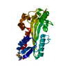
+60S Ribosomal protein ... , 33 types, 33 molecules BaBbBcBdBeBfBhBiBjBkBlBmBnBoBpB7BqBrBsBtBuBvB8BwBxByB9BzB2B3...
-Protein , 1 types, 1 molecules Bg
| #59: Protein | Mass: 34309.418 Da / Num. of mol.: 1 / Source method: isolated from a natural source / Source: (natural) |
|---|
-Details
| Sequence details | OPPOSING STRANDS OF RNA IN SOME EXPANSION SEGMENTS HAVE BEEN NUMBERED CONSECUTIVELY AND COULD ...OPPOSING STRANDS OF RNA IN SOME EXPANSION SEGMENTS HAVE BEEN NUMBERED CONSECUTIV |
|---|
-Experimental details
-Experiment
| Experiment | Method: ELECTRON MICROSCOPY |
|---|---|
| EM experiment | Aggregation state: PARTICLE / 3D reconstruction method: single particle reconstruction |
- Sample preparation
| Component |
| ||||||||||||
|---|---|---|---|---|---|---|---|---|---|---|---|---|---|
| Buffer solution | Name: 30mM Hepes, 50mM KAc, 10mM Mg acetate, 1.5% digitonin / pH: 7.5 Details: 30mM Hepes, 50mM KAc, 10mM Mg acetate, 1.5% digitonin | ||||||||||||
| Specimen | Embedding applied: NO / Shadowing applied: NO / Staining applied: NO / Vitrification applied: YES | ||||||||||||
| Specimen support | Details: thin carbon film on copper 400 mesh grids | ||||||||||||
| Vitrification | Instrument: HOMEMADE PLUNGER / Cryogen name: ETHANE Details: plunge freezing with a custom apparatus at 4C and RH greater than 90 percent |
- Electron microscopy imaging
| Experimental equipment |  Model: Tecnai F20 / Image courtesy: FEI Company |
|---|---|
| Microscopy | Model: FEI TECNAI F20 / Date: Apr 1, 2002 |
| Electron gun | Electron source:  FIELD EMISSION GUN / Accelerating voltage: 200 kV / Illumination mode: FLOOD BEAM FIELD EMISSION GUN / Accelerating voltage: 200 kV / Illumination mode: FLOOD BEAM |
| Electron lens | Mode: BRIGHT FIELD / Nominal magnification: 50000 X / Calibrated magnification: 51000 X / Nominal defocus max: 4400 nm / Nominal defocus min: 1100 nm / Cs: 2 mm |
| Specimen holder | Temperature: 93 K / Tilt angle max: 0 ° / Tilt angle min: 0 ° |
| Image recording | Electron dose: 15 e/Å2 / Film or detector model: KODAK SO-163 FILM |
| Radiation | Protocol: SINGLE WAVELENGTH / Monochromatic (M) / Laue (L): M / Scattering type: x-ray |
| Radiation wavelength | Relative weight: 1 |
- Processing
| EM software |
| ||||||||||||||||||||||||||||||||||||||||||||||||||||||||||||||||||||||
|---|---|---|---|---|---|---|---|---|---|---|---|---|---|---|---|---|---|---|---|---|---|---|---|---|---|---|---|---|---|---|---|---|---|---|---|---|---|---|---|---|---|---|---|---|---|---|---|---|---|---|---|---|---|---|---|---|---|---|---|---|---|---|---|---|---|---|---|---|---|---|---|
| CTF correction | Details: CTF correction using phase flipping and setsf in EMAN to correct the amplitudes | ||||||||||||||||||||||||||||||||||||||||||||||||||||||||||||||||||||||
| Symmetry | Point symmetry: C1 (asymmetric) | ||||||||||||||||||||||||||||||||||||||||||||||||||||||||||||||||||||||
| 3D reconstruction | Method: projection matching / Resolution: 8.7 Å / Num. of particles: 78800 / Nominal pixel size: 2.73 Å / Actual pixel size: 2.73 Å Magnification calibration: Used the 4.6A spacing of vermiculite as a standard Details: Refinements done in EMAN / Symmetry type: POINT | ||||||||||||||||||||||||||||||||||||||||||||||||||||||||||||||||||||||
| Atomic model building | Protocol: RIGID BODY FIT / Space: REAL Target criteria: Rigid body fitting and best visual fit using the program O for RNA. For the proteins DOPE score and other statistical techniques were used to evaluate the best fold and a local ...Target criteria: Rigid body fitting and best visual fit using the program O for RNA. For the proteins DOPE score and other statistical techniques were used to evaluate the best fold and a local exhaustive exploration of Euler angles was used to evaluate the best fit into the density. Details: REFINEMENT PROTOCOL--Rigid body | ||||||||||||||||||||||||||||||||||||||||||||||||||||||||||||||||||||||
| Atomic model building |
| ||||||||||||||||||||||||||||||||||||||||||||||||||||||||||||||||||||||
| Refinement step | Cycle: LAST
|