 Movie
Movie Controller


+ Open data

- Basic information
| Entry | Database: PDB / ID: 5a2q | |||||||||
|---|---|---|---|---|---|---|---|---|---|---|
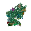
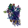
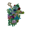
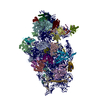
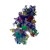
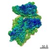
| Title | Structure of the HCV IRES bound to the human ribosome | |||||||||
 Components Components |
| |||||||||
 Keywords Keywords | RIBOSOME / HUMAN RIBOSOME / HEPATITIS-C / IRES / TRANSLATION INITIATION | |||||||||
| Function / homology |  Function and homology information Function and homology informationexit from mitosis / optic nerve development / negative regulation of endoplasmic reticulum unfolded protein response / retinal ganglion cell axon guidance / oxidized pyrimidine DNA binding / response to TNF agonist / positive regulation of base-excision repair / positive regulation of respiratory burst involved in inflammatory response / positive regulation of gastrulation / positive regulation of intrinsic apoptotic signaling pathway in response to DNA damage ...exit from mitosis / optic nerve development / negative regulation of endoplasmic reticulum unfolded protein response / retinal ganglion cell axon guidance / oxidized pyrimidine DNA binding / response to TNF agonist / positive regulation of base-excision repair / positive regulation of respiratory burst involved in inflammatory response / positive regulation of gastrulation / positive regulation of intrinsic apoptotic signaling pathway in response to DNA damage / protein tyrosine kinase inhibitor activity / positive regulation of ubiquitin-protein transferase activity / IRE1-RACK1-PP2A complex / positive regulation of Golgi to plasma membrane protein transport / nucleolus organization / positive regulation of DNA-templated transcription initiation / TNFR1-mediated ceramide production / negative regulation of RNA splicing / neural crest cell differentiation / ZNF598 and the Ribosome-associated Quality Trigger (RQT) complex dissociate a ribosome stalled on a no-go mRNA / supercoiled DNA binding / negative regulation of DNA repair / PELO:HBS1L and ABCE1 dissociate a ribosome on a non-stop mRNA / cysteine-type endopeptidase activator activity involved in apoptotic process / oxidized purine DNA binding / NF-kappaB complex / cytoplasmic translational initiation / rRNA modification in the nucleus and cytosol / negative regulation of intrinsic apoptotic signaling pathway in response to hydrogen peroxide / erythrocyte homeostasis / regulation of establishment of cell polarity / negative regulation of phagocytosis / negative regulation of bicellular tight junction assembly / ubiquitin-like protein conjugating enzyme binding / cytoplasmic side of rough endoplasmic reticulum membrane / Formation of the ternary complex, and subsequently, the 43S complex / pigmentation / ion channel inhibitor activity / protein kinase A binding / laminin receptor activity / Ribosomal scanning and start codon recognition / positive regulation of mitochondrial depolarization / Translation initiation complex formation / negative regulation of Wnt signaling pathway / fibroblast growth factor binding / Protein hydroxylation / BH3 domain binding / negative regulation of translational frameshifting / TOR signaling / regulation of adenylate cyclase-activating G protein-coupled receptor signaling pathway / monocyte chemotaxis / mTORC1-mediated signalling / SARS-CoV-1 modulates host translation machinery / regulation of cell division / iron-sulfur cluster binding / positive regulation of GTPase activity / cellular response to ethanol / Peptide chain elongation / Selenocysteine synthesis / negative regulation of protein binding / Formation of a pool of free 40S subunits / positive regulation of intrinsic apoptotic signaling pathway by p53 class mediator / Eukaryotic Translation Termination / protein serine/threonine kinase inhibitor activity / negative regulation of respiratory burst involved in inflammatory response / SRP-dependent cotranslational protein targeting to membrane / Response of EIF2AK4 (GCN2) to amino acid deficiency / ubiquitin ligase inhibitor activity / Viral mRNA Translation / endonucleolytic cleavage to generate mature 3'-end of SSU-rRNA from (SSU-rRNA, 5.8S rRNA, LSU-rRNA) / Nonsense Mediated Decay (NMD) independent of the Exon Junction Complex (EJC) / positive regulation of signal transduction by p53 class mediator / negative regulation of ubiquitin-dependent protein catabolic process / GTP hydrolysis and joining of the 60S ribosomal subunit / L13a-mediated translational silencing of Ceruloplasmin expression / Major pathway of rRNA processing in the nucleolus and cytosol / regulation of translational fidelity / positive regulation of microtubule polymerization / phagocytic cup / Nonsense Mediated Decay (NMD) enhanced by the Exon Junction Complex (EJC) / spindle assembly / cellular response to leukemia inhibitory factor / positive regulation of intrinsic apoptotic signaling pathway / Protein methylation / endonucleolytic cleavage in ITS1 to separate SSU-rRNA from 5.8S rRNA and LSU-rRNA from tricistronic rRNA transcript (SSU-rRNA, 5.8S rRNA, LSU-rRNA) / translation regulator activity / Nuclear events stimulated by ALK signaling in cancer / gastrulation / rough endoplasmic reticulum / ribosomal small subunit export from nucleus / Amplification of signal from unattached kinetochores via a MAD2 inhibitory signal / laminin binding / DNA-(apurinic or apyrimidinic site) endonuclease activity / positive regulation of cell cycle / rescue of stalled cytosolic ribosome / signaling adaptor activity / Maturation of protein E / negative regulation of protein ubiquitination / Maturation of protein E / MDM2/MDM4 family protein binding Similarity search - Function | |||||||||
| Biological species |  HOMO SAPIENS (human) HOMO SAPIENS (human) HEPATITIS C VIRUS HEPATITIS C VIRUS | |||||||||
| Method | ELECTRON MICROSCOPY / single particle reconstruction / cryo EM / Resolution: 3.9 Å | |||||||||
 Authors Authors | Quade, N. / Leiundgut, M. / Boehringer, D. / Heuvel, J.v.d. / Ban, N. | |||||||||
 Citation Citation | Journal: Nat Commun / Year: 2015 Title: Cryo-EM structure of Hepatitis C virus IRES bound to the human ribosome at 3.9-Å resolution. Authors: Nick Quade / Daniel Boehringer / Marc Leibundgut / Joop van den Heuvel / Nenad Ban /   Abstract: Hepatitis C virus (HCV), a widespread human pathogen, is dependent on a highly structured 5'-untranslated region of its mRNA, referred to as internal ribosome entry site (IRES), for the translation ...Hepatitis C virus (HCV), a widespread human pathogen, is dependent on a highly structured 5'-untranslated region of its mRNA, referred to as internal ribosome entry site (IRES), for the translation of all of its proteins. The HCV IRES initiates translation by directly binding to the small ribosomal subunit (40S), circumventing the need for many eukaryotic translation initiation factors required for mRNA scanning. Here we present the cryo-EM structure of the human 40S ribosomal subunit in complex with the HCV IRES at 3.9 Å resolution, determined by focused refinement of an 80S ribosome-HCV IRES complex. The structure reveals the molecular details of the interactions between the IRES and the 40S, showing that expansion segment 7 (ES7) of the 18S rRNA acts as a central anchor point for the HCV IRES. The structural data rationalizes previous biochemical and genetic evidence regarding the initiation mechanism of the HCV and other related IRESs. | |||||||||
| History |
| |||||||||
| Remark 700 | SHEET DETERMINATION METHOD: DSSP THE SHEETS PRESENTED AS "IC" IN EACH CHAIN ON SHEET RECORDS BELOW ... SHEET DETERMINATION METHOD: DSSP THE SHEETS PRESENTED AS "IC" IN EACH CHAIN ON SHEET RECORDS BELOW IS ACTUALLY AN 5-STRANDED BARREL THIS IS REPRESENTED BY A 6-STRANDED SHEET IN WHICH THE FIRST AND LAST STRANDS ARE IDENTICAL. THE SHEETS PRESENTED AS "LA" IN EACH CHAIN ON SHEET RECORDS BELOW IS ACTUALLY AN 5-STRANDED BARREL THIS IS REPRESENTED BY A 6-STRANDED SHEET IN WHICH THE FIRST AND LAST STRANDS ARE IDENTICAL. THE SHEETS PRESENTED AS "XA" IN EACH CHAIN ON SHEET RECORDS BELOW IS ACTUALLY AN 5-STRANDED BARREL THIS IS REPRESENTED BY A 6-STRANDED SHEET IN WHICH THE FIRST AND LAST STRANDS ARE IDENTICAL. |
- Structure visualization
| Movie |
Movie viewer |
|---|---|
| Structure viewer | Molecule: MolmilJmol/JSmol |
- Downloads & links
-Download
| PDBx/mmCIF format | 5a2q.cif.gz | 1.8 MB | Display | PDBx/mmCIF format |
|---|---|---|---|---|
| PDB format | pdb5a2q.ent.gz | 1.4 MB | Display | PDB format |
| PDBx/mmJSON format | 5a2q.json.gz | Tree view | PDBx/mmJSON format | |
| Others |  Other downloads Other downloads |
-Validation report
| Arichive directory | https://data.pdbj.org/pub/pdb/validation_reports/a2/5a2qftp://data.pdbj.org/pub/pdb/validation_reports/a2/5a2q | HTTPS FTP |
|---|
-Related structure data
| Related structure data | 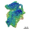 3019MC M: map data used to model this data C: citing same article ( |
|---|---|
| Similar structure data |


-Links
 PDBj
PDBj


- Assembly
| Deposited unit | 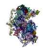
|
|---|---|
| 1 |
|
-Components
-RNA chain , 2 types, 2 molecules 23
| #1: RNA chain | Mass: 602432.625 Da / Num. of mol.: 1 / Source method: isolated from a natural source / Source: (natural) HOMO SAPIENS (human) / Cell line: HEK293-6E / Organ: KIDNEY / References: GenBank: 337376 |
|---|---|
| #2: RNA chain | Mass: 82914.953 Da / Num. of mol.: 1 / Source method: obtained synthetically / Source: (synth.) HEPATITIS C VIRUS |
+RIBOSOMAL PROTEIN ... , 36 types, 36 molecules ABCDEFGHIJKLMNOPQRSTUVWXYZabcd...
-Non-polymers , 3 types, 245 molecules 


| #39: Chemical | ChemComp-MG /  Mass: 24.305 Da / Num. of mol.: 98 / Source method: obtained synthetically / Formula: Mg Mass: 24.305 Da / Num. of mol.: 98 / Source method: obtained synthetically / Formula: Mg#40: Chemical |  Mass: 65.409 Da / Num. of mol.: 3 / Source method: obtained synthetically / Formula: Zn Mass: 65.409 Da / Num. of mol.: 3 / Source method: obtained synthetically / Formula: Zn#41: Water | ChemComp-HOH / | Mass: 18.015 Da / Num. of mol.: 144 / Source method: isolated from a natural source / Formula: H2O |
|---|
-Details
| Has protein modification | Y |
|---|---|
| Sequence details | GENBANK REFERENCE FOR CHAIN A CAA54808.1 GENBANK REFERENCE FOR CHAIN G AAH27620.1 |
-Experimental details
-Experiment
| Experiment | Method: ELECTRON MICROSCOPY |
|---|---|
| EM experiment | Aggregation state: PARTICLE / 3D reconstruction method: single particle reconstruction |
- Sample preparation
| Component | Name: HCV IRES BOUND TO HUMAN 80S RIBOSOME / Type: RIBOSOME |
|---|---|
| Buffer solution | Name: 20MM HEPES, 100 MM KCL, 5 MM MGCL2 / pH: 7.6 / Details: 20MM HEPES, 100 MM KCL, 5 MM MGCL2 |
| Specimen | Conc.: 0.4 mg/ml / Embedding applied: NO / Shadowing applied: NO / Staining applied: NO / Vitrification applied: YES |
| Specimen support | Details: HOLEY CARBON |
| Vitrification | Instrument: HOMEMADE PLUNGER / Cryogen name: ETHANE-PROPANE Details: FROZEN ON 200 MESH QUANTIFOIL R 2 2 HOLEY CARBON GRIDS WITH A THIN CONTINUOUS CARBON SUPPORT FILM APPLIED IN ETHANE PROPANE MIX |
- Electron microscopy imaging
| Experimental equipment |  Model: Titan Krios / Image courtesy: FEI Company |
|---|---|
| Microscopy | Model: FEI TITAN KRIOS / Date: Nov 20, 2014 |
| Electron gun | Electron source:  FIELD EMISSION GUN / Accelerating voltage: 300 kV / Illumination mode: FLOOD BEAM FIELD EMISSION GUN / Accelerating voltage: 300 kV / Illumination mode: FLOOD BEAM |
| Electron lens | Mode: BRIGHT FIELD / Nominal magnification: 100719 X / Nominal defocus max: 3400 nm / Nominal defocus min: 1500 nm / Cs: 2.7 mm |
| Image recording | Electron dose: 20 e/Å2 / Film or detector model: FEI FALCON II (4k x 4k) |
| Radiation wavelength | Relative weight: 1 |
- Processing
| EM software |
| ||||||||||||
|---|---|---|---|---|---|---|---|---|---|---|---|---|---|
| CTF correction | Details: INDIVIDUAL FRAMES | ||||||||||||
| Symmetry | Point symmetry: C1 (asymmetric) | ||||||||||||
| 3D reconstruction | Method: MAXIMUM LIKELIHOOD BASED REFINEMENT / Resolution: 3.9 Å / Num. of particles: 404357 / Nominal pixel size: 1.39 Å / Symmetry type: POINT | ||||||||||||
| Atomic model building | B value: 119.1 / Protocol: RIGID BODY FIT / Space: REAL / Target criteria: R-factor Details: METHOD--RIGID BODY REFINEMENT FOLLOWED BY MANUAL MODEL BUILDING REFINEMENT PROTOCOL--CRYO-EM | ||||||||||||
| Atomic model building | PDB-ID: 4W23 4w23 Accession code: 4W23 / Source name: PDB / Type: experimental model | ||||||||||||
| Refinement | Highest resolution: 3.9 Å | ||||||||||||
| Refinement step | Cycle: LAST / Highest resolution: 3.9 Å
|
