 Movie
Movie Controller

[English] 日本語
 Yorodumi
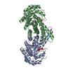
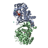
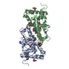
- PDB-4d48: Crystal Structure of glucose-1-phosphate uridylyltransferase GalU... -
+ Open data

- Basic information
| Entry | Database: PDB / ID: 4d48 | ||||||
|---|---|---|---|---|---|---|---|
| Title | Crystal Structure of glucose-1-phosphate uridylyltransferase GalU from Erwinia amylovora. | ||||||
 Components Components | GLUCOSE-1-PHOSPHATE URIDYLYLTRANSFERASE | ||||||
 Keywords Keywords | TRANSFERASE / AMYLOVORAN BIOSYNTHESIS / UDP-GLUCOSE / FIRE BLIGHT | ||||||
| Function / homology |  Function and homology information Function and homology informationUTP-glucose-1-phosphate uridylyltransferase / UTP:glucose-1-phosphate uridylyltransferase activity / UDP-alpha-D-glucose metabolic process / biosynthetic process Similarity search - Function | ||||||
| Biological species |  ERWINIA AMYLOVORA (bacteria) ERWINIA AMYLOVORA (bacteria) | ||||||
| Method |  X-RAY DIFFRACTION / SYNCHROTRON / MOLECULAR REPLACEMENT / Resolution: 2.46 Å X-RAY DIFFRACTION / SYNCHROTRON / MOLECULAR REPLACEMENT / Resolution: 2.46 Å | ||||||
 Authors Authors | Toccafondi, M. / Wuerges, J. / Cianci, M. / Benini, S. | ||||||
 Citation Citation | Journal: Biochim. Biophys. Acta / Year: 2017 Title: Glucose-1-phosphate uridylyltransferase from Erwinia amylovora: Activity, structure and substrate specificity. Authors: Benini, S. / Toccafondi, M. / Rejzek, M. / Musiani, F. / Wagstaff, B.A. / Wuerges, J. / Cianci, M. / Field, R.A. #1: Journal: Acta Crystallogr F Struct Biol Commun / Year: 2014 Title: Expression, purification, crystallization and preliminary X-ray analysis of glucose-1-phosphate uridylyltransferase (GalU) from Erwinia amylovora. Authors: Toccafondi, M. / Cianci, M. / Benini, S. | ||||||
| History |
|
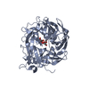
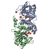
- Structure visualization
| Structure viewer | Molecule: MolmilJmol/JSmol |
|---|
- Downloads & links
-Download
| PDBx/mmCIF format | 4d48.cif.gz | 228.3 KB | Display | PDBx/mmCIF format |
|---|---|---|---|---|
| PDB format | pdb4d48.ent.gz | 187 KB | Display | PDB format |
| PDBx/mmJSON format | 4d48.json.gz | Tree view | PDBx/mmJSON format | |
| Others |  Other downloads Other downloads |
-Validation report
| Arichive directory | https://data.pdbj.org/pub/pdb/validation_reports/d4/4d48ftp://data.pdbj.org/pub/pdb/validation_reports/d4/4d48 | HTTPS FTP |
|---|
-Related structure data
| Related structure data | 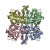 2e3dS S: Starting model for refinement |
|---|---|
| Similar structure data |


-Links
 PDBj
PDBj

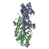
- Assembly
| Deposited unit | 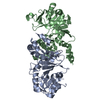
| ||||||||
|---|---|---|---|---|---|---|---|---|---|
| 1 |
| ||||||||
| Unit cell |
| ||||||||
| Noncrystallographic symmetry (NCS) | NCS oper: (Code: given Matrix: (0.466, 0.8848, -0.001262), Vector: |
-Components
| #1: Protein | Mass: 32922.973 Da / Num. of mol.: 2 Source method: isolated from a genetically manipulated source Source: (gene. exp.) ERWINIA AMYLOVORA (bacteria) / Strain: CFBP1430 / Production host: References: UniProt: D4I3X5, UTP-glucose-1-phosphate uridylyltransferase #2: Water | ChemComp-HOH / |  Mass: 18.015 Da / Num. of mol.: 14 / Source method: isolated from a natural source / Formula: H2O Mass: 18.015 Da / Num. of mol.: 14 / Source method: isolated from a natural source / Formula: H2O |
|---|
-Experimental details
-Experiment
| Experiment | Method: X-RAY DIFFRACTION / Number of used crystals: 1 |
|---|
- Sample preparation
| Crystal | Density Matthews: 2.57 Å3/Da / Density % sol: 52.09 % / Description: NONE |
|---|---|
| Crystal grow | pH: 8.5 Details: 2M AMMONIUM SULFATE, 0.1 M TRIS PH 8.5, 5% ETHYLENE GLYCOL |
-Data collection
| Diffraction | Mean temperature: 100 K |
|---|---|
| Diffraction source | Source: SYNCHROTRON / Site: PETRA III, EMBL c/o DESY  / Beamline: P13 (MX1) / Wavelength: 1.033 / Beamline: P13 (MX1) / Wavelength: 1.033 |
| Detector | Type: DECTRIS PILATUS 6M / Detector: PIXEL / Date: Jan 10, 2014 |
| Radiation | Monochromator: SI(111) / Protocol: SINGLE WAVELENGTH / Monochromatic (M) / Laue (L): M / Scattering type: x-ray |
| Radiation wavelength | Wavelength: 1.033 Å / Relative weight: 1 |
| Reflection | Resolution: 2.46→66.66 Å / Num. obs: 12434 / % possible obs: 99.4 % / Observed criterion σ(I): 3 / Redundancy: 8.7 % / Biso Wilson estimate: 57.06 Å2 / Rmerge(I) obs: 0.1 / Net I/σ(I): 12.1 |
| Reflection shell | Resolution: 2.46→2.56 Å / Redundancy: 5.6 % / Rmerge(I) obs: 0.43 / Mean I/σ(I) obs: 3.4 / % possible all: 94.9 |
- Processing
| Software |
| ||||||||||||||||||||||||||||||||||||||||||||||||||||||||||||||||||||||||||||||||||||||||||||||||||||||||||||||||||||||||||||||||||||||||||||||||||||||||||||||||||||||||||||||||||||||
|---|---|---|---|---|---|---|---|---|---|---|---|---|---|---|---|---|---|---|---|---|---|---|---|---|---|---|---|---|---|---|---|---|---|---|---|---|---|---|---|---|---|---|---|---|---|---|---|---|---|---|---|---|---|---|---|---|---|---|---|---|---|---|---|---|---|---|---|---|---|---|---|---|---|---|---|---|---|---|---|---|---|---|---|---|---|---|---|---|---|---|---|---|---|---|---|---|---|---|---|---|---|---|---|---|---|---|---|---|---|---|---|---|---|---|---|---|---|---|---|---|---|---|---|---|---|---|---|---|---|---|---|---|---|---|---|---|---|---|---|---|---|---|---|---|---|---|---|---|---|---|---|---|---|---|---|---|---|---|---|---|---|---|---|---|---|---|---|---|---|---|---|---|---|---|---|---|---|---|---|---|---|---|---|
| Refinement | Method to determine structure: MOLECULAR REPLACEMENT Starting model: PDB ENTRY 2E3D Resolution: 2.46→64.66 Å / Cor.coef. Fo:Fc: 0.923 / Cor.coef. Fo:Fc free: 0.909 / SU B: 28.167 / SU ML: 0.298 / Cross valid method: THROUGHOUT / ESU R: 0.658 / ESU R Free: 0.311 / Stereochemistry target values: MAXIMUM LIKELIHOOD Details: HYDROGENS HAVE BEEN ADDED IN THE RIDING POSITIONS. U VALUES WITH TLS ADDED. THE FOLLOWING RESIDUES WERE DISORDERED 1-4, 84-87, 233-238, 299-302
| ||||||||||||||||||||||||||||||||||||||||||||||||||||||||||||||||||||||||||||||||||||||||||||||||||||||||||||||||||||||||||||||||||||||||||||||||||||||||||||||||||||||||||||||||||||||
| Solvent computation | Ion probe radii: 0.8 Å / Shrinkage radii: 0.8 Å / VDW probe radii: 1.2 Å / Solvent model: MASK | ||||||||||||||||||||||||||||||||||||||||||||||||||||||||||||||||||||||||||||||||||||||||||||||||||||||||||||||||||||||||||||||||||||||||||||||||||||||||||||||||||||||||||||||||||||||
| Displacement parameters | Biso mean: 70.542 Å2
| ||||||||||||||||||||||||||||||||||||||||||||||||||||||||||||||||||||||||||||||||||||||||||||||||||||||||||||||||||||||||||||||||||||||||||||||||||||||||||||||||||||||||||||||||||||||
| Refinement step | Cycle: LAST / Resolution: 2.46→64.66 Å
| ||||||||||||||||||||||||||||||||||||||||||||||||||||||||||||||||||||||||||||||||||||||||||||||||||||||||||||||||||||||||||||||||||||||||||||||||||||||||||||||||||||||||||||||||||||||
| Refine LS restraints |
|