 Movie
Movie Controller

[English] 日本語
 Yorodumi
- PDB-7nc7: Crystal structure of fructose-bisphosphate aldolases FBAC from Ba... -
+ Open data

- Basic information
| Entry | Database: PDB / ID: 7nc7 | ||||||
|---|---|---|---|---|---|---|---|
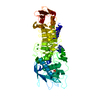
| Title | Crystal structure of fructose-bisphosphate aldolases FBAC from Bacillus methanolicus | ||||||
 Components Components | Fructose-bisphosphate aldolase | ||||||
 Keywords Keywords | LYASE / glycolytic aldolase | ||||||
| Function / homology |  Function and homology information Function and homology informationfructose-bisphosphate aldolase / fructose-bisphosphate aldolase activity / fructose 1,6-bisphosphate metabolic process / glycolytic process / zinc ion binding Similarity search - Function | ||||||
| Biological species |  | ||||||
| Method |  X-RAY DIFFRACTION / MOLECULAR REPLACEMENT / Resolution: 2.2 Å X-RAY DIFFRACTION / MOLECULAR REPLACEMENT / Resolution: 2.2 Å | ||||||
 Authors Authors | Einsle, O. / Zhang, L. / Guetle, D. / Jacquot, J.P. | ||||||
 Citation Citation | Journal: Front Microbiol / Year: 2021 Title: Interrogating the Role of the Two Distinct Fructose-Bisphosphate Aldolases of Bacillus methanolicus by Site-Directed Mutagenesis of Key Amino Acids and Gene Repression by CRISPR Interference Authors: Schultenkamper, K. / Gutle, D.D. / Lopez, M.G. / Keller, L.B. / Zhang, L. / Einsle, O. / Jacquot, J.P. / Wendisch, V.F. | ||||||
| History |
|
- Structure visualization
| Structure viewer | Molecule: MolmilJmol/JSmol |
|---|
- Downloads & links
-Download
| PDBx/mmCIF format | 7nc7.cif.gz | 117.8 KB | Display | PDBx/mmCIF format |
|---|---|---|---|---|
| PDB format | pdb7nc7.ent.gz | 91.9 KB | Display | PDB format |
| PDBx/mmJSON format | 7nc7.json.gz | Tree view | PDBx/mmJSON format | |
| Others |  Other downloads Other downloads |
-Validation report
| Arichive directory | https://data.pdbj.org/pub/pdb/validation_reports/nc/7nc7ftp://data.pdbj.org/pub/pdb/validation_reports/nc/7nc7 | HTTPS FTP |
|---|
-Related structure data
| Related structure data |  7nccC  3q94S S: Starting model for refinement C: citing same article ( |
|---|---|
| Similar structure data |


-Links
 PDBj
PDBj
- Assembly
| Deposited unit | 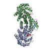
| ||||||||
|---|---|---|---|---|---|---|---|---|---|
| 1 |
| ||||||||
| Unit cell |
|
-Components
| #1: Protein | Mass: 30742.129 Da / Num. of mol.: 2 Source method: isolated from a genetically manipulated source Source: (gene. exp.) Strain: MGA3 / ATCC 53907 / Gene: fbaC, BMMGA3_16125 / Production host: #2: Chemical |   Mass: 170.058 Da / Num. of mol.: 2 / Source method: obtained synthetically / Formula: C3H7O6P / Feature type: SUBJECT OF INVESTIGATION Mass: 170.058 Da / Num. of mol.: 2 / Source method: obtained synthetically / Formula: C3H7O6P / Feature type: SUBJECT OF INVESTIGATION#3: Water | ChemComp-HOH / |  Mass: 18.015 Da / Num. of mol.: 150 / Source method: isolated from a natural source / Formula: H2O Mass: 18.015 Da / Num. of mol.: 150 / Source method: isolated from a natural source / Formula: H2OHas ligand of interest | Y | |
|---|
-Experimental details
-Experiment
| Experiment | Method: X-RAY DIFFRACTION / Number of used crystals: 1 |
|---|
- Sample preparation
| Crystal | Density Matthews: 2 Å3/Da / Density % sol: 38.62 % |
|---|---|
| Crystal grow | Temperature: 293 K / Method: vapor diffusion, sitting drop Details: 0.1 M sodium acetate trihydrate pH 4.5, 2.0 M ammonium sulfate |
-Data collection
| Diffraction | Mean temperature: 100 K / Serial crystal experiment: N |
|---|---|
| Diffraction source | Source: ROTATING ANODE / Type: RIGAKU MICROMAX-007 HF / Wavelength: 1.5418 Å |
| Detector | Type: MAR scanner 345 mm plate / Detector: IMAGE PLATE / Date: Jul 31, 2014 |
| Radiation | Protocol: SINGLE WAVELENGTH / Monochromatic (M) / Laue (L): M / Scattering type: x-ray |
| Radiation wavelength | Wavelength: 1.5418 Å / Relative weight: 1 |
| Reflection | Resolution: 2.2→27.42 Å / Num. obs: 24421 / % possible obs: 99.8 % / Redundancy: 4.1 % / CC1/2: 0.996 / Net I/σ(I): 6.7 |
| Reflection shell | Resolution: 2.2→2.28 Å / Num. unique obs: 2158 / CC1/2: 0.521 |
- Processing
| Software |
| ||||||||||||||||||||||||||||||||||||||||||||||||||||||||||||
|---|---|---|---|---|---|---|---|---|---|---|---|---|---|---|---|---|---|---|---|---|---|---|---|---|---|---|---|---|---|---|---|---|---|---|---|---|---|---|---|---|---|---|---|---|---|---|---|---|---|---|---|---|---|---|---|---|---|---|---|---|---|
| Refinement | Method to determine structure: MOLECULAR REPLACEMENT Starting model: 3Q94 Resolution: 2.2→27.42 Å / Cor.coef. Fo:Fc: 0.953 / Cor.coef. Fo:Fc free: 0.913 / SU B: 9.398 / SU ML: 0.227 / Cross valid method: THROUGHOUT / σ(F): 0 / ESU R Free: 0.319 / Stereochemistry target values: MAXIMUM LIKELIHOOD Details: HYDROGENS HAVE BEEN ADDED IN THE RIDING POSITIONS U VALUES : REFINED INDIVIDUALLY
| ||||||||||||||||||||||||||||||||||||||||||||||||||||||||||||
| Solvent computation | Ion probe radii: 0.8 Å / Shrinkage radii: 0.8 Å / VDW probe radii: 1.2 Å / Solvent model: MASK | ||||||||||||||||||||||||||||||||||||||||||||||||||||||||||||
| Displacement parameters | Biso max: 155.81 Å2 / Biso mean: 36.602 Å2 / Biso min: 17.22 Å2
| ||||||||||||||||||||||||||||||||||||||||||||||||||||||||||||
| Refinement step | Cycle: final / Resolution: 2.2→27.42 Å
| ||||||||||||||||||||||||||||||||||||||||||||||||||||||||||||
| Refine LS restraints |
| ||||||||||||||||||||||||||||||||||||||||||||||||||||||||||||
| LS refinement shell | Resolution: 2.2→2.257 Å / Rfactor Rfree error: 0 / Total num. of bins used: 20
|
