- PDB-6rqa: Crystal structure of the iminosuccinate reductase of Paracoccus d... -
+
Open data
ID or keywords:
Loading...
-
Basic information
Entry
Database: PDB / ID: 6rqa
Title
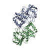
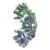
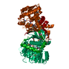
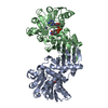
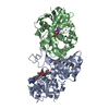
Crystal structure of the iminosuccinate reductase of Paracoccus denitrificans in complex with NAD+
Components
iminosuccinate reductase
Keywords
OXIDOREDUCTASE / iminosuccinate reductase
Function / homology
Function and homology information
Oxidoreductases; Acting on the CH-NH2 group of donors; With NAD+ or NADP+ as acceptor / oxidoreductase activity, acting on the CH-NH2 group of donors, NAD or NADP as acceptor / glycolate catabolic process / glyoxylate catabolic process / NADH binding / cytoplasm Similarity search - Function
Mass: 18.015 Da / Num. of mol.: 127 / Source method: isolated from a natural source / Formula: H2O
-
Experimental details
-
Experiment
Experiment
Method: X-RAY DIFFRACTION / Number of used crystals: 1
-
Sample preparation
Crystal
Density Matthews: 2.2 Å3/Da / Density % sol: 44.19 % / Description: thick plates
Crystal grow
Temperature: 289 K / Method: vapor diffusion, sitting drop / pH: 5.9 Details: His-tagged iminosuccinate reductase (10 mg/ml) in buffer containing 25 mM Tris-HCl (pH 8.0), 100 mM NaCl, 1 mM MgCl2, 0.1 mM DTT, 5 mM Tb-Xo4, and 5 mM NAD+ was mixed in a ratio of 1:1 with ...Details: His-tagged iminosuccinate reductase (10 mg/ml) in buffer containing 25 mM Tris-HCl (pH 8.0), 100 mM NaCl, 1 mM MgCl2, 0.1 mM DTT, 5 mM Tb-Xo4, and 5 mM NAD+ was mixed in a ratio of 1:1 with crystallization buffer containing 200 mM Mg(NO3)2 and 20% (w/v) PEG3350 (pH 5.9).
-
Data collection
Diffraction
Mean temperature: 100 K / Serial crystal experiment: N
Diffraction source
Source: SYNCHROTRON / Site: PETRA III, EMBL c/o DESY / Beamline: P13 (MX1) / Wavelength: 0.97625 Å
In the structure databanks used in Yorodumi, some data are registered as the other names, "COVID-19 virus" and "2019-nCoV". Here are the details of the virus and the list of structure data.
Jan 31, 2019. EMDB accession codes are about to change! (news from PDBe EMDB page)
EMDB accession codes are about to change! (news from PDBe EMDB page)
The allocation of 4 digits for EMDB accession codes will soon come to an end. Whilst these codes will remain in use, new EMDB accession codes will include an additional digit and will expand incrementally as the available range of codes is exhausted. The current 4-digit format prefixed with “EMD-” (i.e. EMD-XXXX) will advance to a 5-digit format (i.e. EMD-XXXXX), and so on. It is currently estimated that the 4-digit codes will be depleted around Spring 2019, at which point the 5-digit format will come into force.
The EM Navigator/Yorodumi systems omit the EMD- prefix.
Related info.:Q: What is EMD? / ID/Accession-code notation in Yorodumi/EM Navigator
Yorodumi is a browser for structure data from EMDB, PDB, SASBDB, etc.
This page is also the successor to EM Navigator detail page, and also detail information page/front-end page for Omokage search.
The word "yorodu" (or yorozu) is an old Japanese word meaning "ten thousand". "mi" (miru) is to see.
Related info.:EMDB / PDB / SASBDB / Comparison of 3 databanks / Yorodumi Search / Aug 31, 2016. New EM Navigator & Yorodumi / Yorodumi Papers / Jmol/JSmol / Function and homology information / Changes in new EM Navigator and Yorodumi
 Movie
Movie Controller

 Yorodumi
 Open data
 Basic information
 Components
 Keywords
 Function and homology information
 Paracoccus denitrificans (bacteria)
 X-RAY DIFFRACTION /
  Authors
 Germany, 2items
  Citation
 Structure visualization
 Downloads & links
 Other downloads
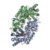


 PDBj
PDBj Assembly
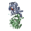

 Mass: 663.425 Da / Num. of mol.: 2 / Source method: obtained synthetically / Formula: C21H27N7O14P2 / Feature type: SUBJECT OF INVESTIGATION / Comment: NAD*YM
 Mass: 158.925 Da / Num. of mol.: 2 / Source method: obtained synthetically / Formula: Tb
 Mass: 556.353 Da / Num. of mol.: 1 / Source method: obtained synthetically / Formula: C20H23N5O4Tb
 Mass: 18.015 Da / Num. of mol.: 127 / Source method: isolated from a natural source / Formula: H2O
 Sample preparation
 Processing