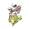
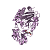
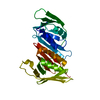
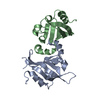
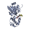
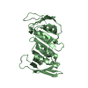
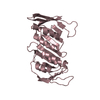
- PDB-4cs5: Crystal Structure of PCNA from Litopenaeus vannamei -
+
Open data
ID or keywords:
Loading...
-
Basic information
Entry
Database: PDB / ID: 4cs5
Title
Crystal Structure of PCNA from Litopenaeus vannamei
Components
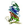
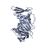
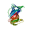
PROLIFERATING CELL NUCLEAR ANTIGEN
Keywords
NUCLEAR PROTEIN / DNA BINDING PROTEIN
Function / homology
Function and homology information
PCNA complex / leading strand elongation / DNA polymerase processivity factor activity / regulation of DNA replication / mismatch repair / translesion synthesis / DNA binding Similarity search - Function
CHAINAAND (RESSEQ1:254 ) AND (NOTELEMENTH) AND (NOTELEMENTD)
2
1
1
CHAINBAND (RESSEQ1:254 ) AND (NOTELEMENTH) AND (NOTELEMENTD)
3
1
1
CHAINCAND (RESSEQ1:254 ) AND (NOTELEMENTH) AND (NOTELEMENTD)
-
Components
#1: Protein
PROLIFERATINGCELLNUCLEARANTIGEN
Mass: 28840.066 Da / Num. of mol.: 3 Source method: isolated from a genetically manipulated source Source: (gene. exp.) LITOPENAEUS VANNAMEI (Pacific white shrimp) Plasmid: PJ404 / Production host: ESCHERICHIA COLI (E. coli) / Strain (production host): BL21 / Variant (production host): SI / References: UniProt: G1E6N7
Sequence details
THE AMINOACID SEQUENCE OF THE PDB FILE LACK THE LAST 6 C- TERMINAL RESIDUES COMPARED TO THE ...THE AMINOACID SEQUENCE OF THE PDB FILE LACK THE LAST 6 C- TERMINAL RESIDUES COMPARED TO THE SEQUENCE REPORTED IN UNIPROT. THIS IS BECAUSE THERE IS NO ELECTRONIC DENSITY TO INCLUDE THAT RESIDUES IN THE MODEL.
-
Experimental details
-
Experiment
Experiment
Method: X-RAY DIFFRACTION / Number of used crystals: 1
-
Sample preparation
Crystal
Density Matthews: 2.36 Å3/Da / Density % sol: 47.35 %
Crystal grow
pH: 7.5 Details: 300 MM CACL2, 100 MM SODIUM HEPES PH 7.5 AND 30%(V/V) PEG 400
In the structure databanks used in Yorodumi, some data are registered as the other names, "COVID-19 virus" and "2019-nCoV". Here are the details of the virus and the list of structure data.
Jan 31, 2019. EMDB accession codes are about to change! (news from PDBe EMDB page)
EMDB accession codes are about to change! (news from PDBe EMDB page)
The allocation of 4 digits for EMDB accession codes will soon come to an end. Whilst these codes will remain in use, new EMDB accession codes will include an additional digit and will expand incrementally as the available range of codes is exhausted. The current 4-digit format prefixed with “EMD-” (i.e. EMD-XXXX) will advance to a 5-digit format (i.e. EMD-XXXXX), and so on. It is currently estimated that the 4-digit codes will be depleted around Spring 2019, at which point the 5-digit format will come into force.
The EM Navigator/Yorodumi systems omit the EMD- prefix.
Related info.:Q: What is EMD? / ID/Accession-code notation in Yorodumi/EM Navigator
Yorodumi is a browser for structure data from EMDB, PDB, SASBDB, etc.
This page is also the successor to EM Navigator detail page, and also detail information page/front-end page for Omokage search.
The word "yorodu" (or yorozu) is an old Japanese word meaning "ten thousand". "mi" (miru) is to see.
Related info.:EMDB / PDB / SASBDB / Comparison of 3 databanks / Yorodumi Search / Aug 31, 2016. New EM Navigator & Yorodumi / Yorodumi Papers / Jmol/JSmol / Function and homology information / Changes in new EM Navigator and Yorodumi
 Movie
Movie Controller


 Open data
 Basic information
 Components
 Keywords
 Function and homology information
 LITOPENAEUS VANNAMEI (Pacific white shrimp)
 X-RAY DIFFRACTION /
  Authors
 Citation
 Structure visualization
 Downloads & links
 Other downloads
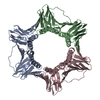


 PDBj
PDBj

 Assembly


 Sample preparation
 / Beamline: X4C / Wavelength: 0.9795
  Processing