 Movie
Movie Controller

[English] 日本語
 Yorodumi
- PDB-1plr: CRYSTAL STRUCTURE OF THE EUKARYOTIC DNA POLYMERASE PROCESSIVITY F... -
+ Open data

- Basic information
| Entry | Database: PDB / ID: 1plr | ||||||
|---|---|---|---|---|---|---|---|
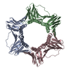
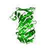
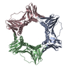
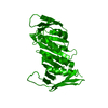
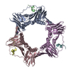
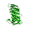
| Title | CRYSTAL STRUCTURE OF THE EUKARYOTIC DNA POLYMERASE PROCESSIVITY FACTOR PCNA | ||||||
 Components Components | PROLIFERATING CELL NUCLEAR ANTIGEN (PCNA) | ||||||
 Keywords Keywords | DNA-BINDING / NUCLEAR PROTEIN / DNA REPLICATION | ||||||
| Function / homology |  Function and homology information Function and homology informationMismatch repair (MMR) directed by MSH2:MSH6 (MutSalpha) / positive regulation of DNA metabolic process / meiotic mismatch repair / Processive synthesis on the lagging strand / Removal of the Flap Intermediate / : / Polymerase switching / maintenance of DNA trinucleotide repeats / Translesion synthesis by REV1 / : ...Mismatch repair (MMR) directed by MSH2:MSH6 (MutSalpha) / positive regulation of DNA metabolic process / meiotic mismatch repair / Processive synthesis on the lagging strand / Removal of the Flap Intermediate / : / Polymerase switching / maintenance of DNA trinucleotide repeats / Translesion synthesis by REV1 / : / : / : / SUMOylation of DNA replication proteins / establishment of mitotic sister chromatid cohesion / PCNA complex / : / lagging strand elongation / DNA damage tolerance / silent mating-type cassette heterochromatin formation / mitotic sister chromatid cohesion / error-free translesion synthesis / leading strand elongation / DNA polymerase processivity factor activity / Dual incision in TC-NER / subtelomeric heterochromatin formation / mismatch repair / translesion synthesis / positive regulation of DNA replication / replication fork / positive regulation of DNA repair / nucleotide-excision repair / mitotic cell cycle / chromosome, telomeric region / DNA binding / identical protein binding / nucleus Similarity search - Function | ||||||
| Biological species |  | ||||||
| Method |  X-RAY DIFFRACTION / Resolution: 3 Å X-RAY DIFFRACTION / Resolution: 3 Å | ||||||
 Authors Authors | Krishna, T.S.R. / Kong, X.-P. / Gary, S. / Burgers, P.M. / Kuriyan, J. | ||||||
 Citation Citation | Journal: Cell(Cambridge,Mass.) / Year: 1994 Title: Crystal structure of the eukaryotic DNA polymerase processivity factor PCNA. Authors: Krishna, T.S. / Kong, X.P. / Gary, S. / Burgers, P.M. / Kuriyan, J. #1: Journal: J.Mol.Biol. / Year: 1994Title: Crystallization of Proliferating Cell Nuclear Antigen (PCNA) from Saccharomyces Cerevisiae Authors: Krishna, T.S.R. / Kong, X.-P. / Gary, S. / Burgers, P. / Kuriyan, J. #2: Journal: J.Mol.Biol. / Year: 1993Title: Sliding Clamps of DNA Polymerases Authors: Kuriyan, J. / Donnell, M. #3: Journal: Cell(Cambridge,Mass.) / Year: 1992Title: Three-Dimensional Structure of the Beta Subunit of E. Coli DNA Polymerase III Holoenzyme: A Sliding DNA Clamp Authors: Kong, X.-P. / Onrust, R. / Donnell, M. / Kuriyan, J. | ||||||
| History |
|
- Structure visualization
| Structure viewer | Molecule: MolmilJmol/JSmol |
|---|
- Downloads & links
-Download
| PDBx/mmCIF format | 1plr.cif.gz | 61.2 KB | Display | PDBx/mmCIF format |
|---|---|---|---|---|
| PDB format | pdb1plr.ent.gz | 45.8 KB | Display | PDB format |
| PDBx/mmJSON format | 1plr.json.gz | Tree view | PDBx/mmJSON format | |
| Others |  Other downloads Other downloads |
-Validation report
| Arichive directory | https://data.pdbj.org/pub/pdb/validation_reports/pl/1plrftp://data.pdbj.org/pub/pdb/validation_reports/pl/1plr | HTTPS FTP |
|---|
-Related structure data


-Links
 PDBj
PDBj


- Assembly
| Deposited unit | 
| ||||||||
|---|---|---|---|---|---|---|---|---|---|
| 1 | 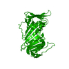
| ||||||||
| Unit cell |
|
-Components
| #1: Protein | Mass: 28944.051 Da / Num. of mol.: 1 Source method: isolated from a genetically manipulated source Source: (gene. exp.) References: UniProt: P15873 |
|---|---|
| #2: Water | ChemComp-HOH /  Mass: 18.015 Da / Num. of mol.: 10 / Source method: isolated from a natural source / Formula: H2O Mass: 18.015 Da / Num. of mol.: 10 / Source method: isolated from a natural source / Formula: H2O |
| Has protein modification | Y |
-Experimental details
-Experiment
| Experiment | Method: X-RAY DIFFRACTION |
|---|
- Sample preparation
| Crystal | Density Matthews: 5.19 Å3/Da / Density % sol: 76.3 % | ||||||||||||||||||||||||||||||||||||
|---|---|---|---|---|---|---|---|---|---|---|---|---|---|---|---|---|---|---|---|---|---|---|---|---|---|---|---|---|---|---|---|---|---|---|---|---|---|
| Crystal grow | *PLUS pH: 7.5 / Method: unknown / Details: Krishna, T.S.R., (1994) J.Mol.Biol., 241, 265. | ||||||||||||||||||||||||||||||||||||
| Components of the solutions | *PLUS
|
-Data collection
| Radiation | Scattering type: x-ray |
|---|---|
| Radiation wavelength | Relative weight: 1 |
| Reflection | Num. obs: 12273 / % possible obs: 86.3 % / Observed criterion σ(I): 1 |
| Reflection | *PLUS Highest resolution: 3 Å / Lowest resolution: 30 Å / Redundancy: 7.8 % / Num. measured all: 95433 / Rmerge(I) obs: 0.065 |
| Reflection shell | *PLUS Highest resolution: 3 Å / Lowest resolution: 3.05 Å / % possible obs: 80.9 % |
- Processing
| Software |
| ||||||||||||||||||||||||||||||||||||||||||||||||||||||||||||
|---|---|---|---|---|---|---|---|---|---|---|---|---|---|---|---|---|---|---|---|---|---|---|---|---|---|---|---|---|---|---|---|---|---|---|---|---|---|---|---|---|---|---|---|---|---|---|---|---|---|---|---|---|---|---|---|---|---|---|---|---|---|
| Refinement | Resolution: 3→6 Å / σ(F): 2 /
| ||||||||||||||||||||||||||||||||||||||||||||||||||||||||||||
| Displacement parameters | Biso mean: 32 Å2 | ||||||||||||||||||||||||||||||||||||||||||||||||||||||||||||
| Refinement step | Cycle: LAST / Resolution: 3→6 Å
| ||||||||||||||||||||||||||||||||||||||||||||||||||||||||||||
| Refine LS restraints |
|
