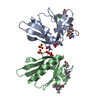
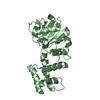
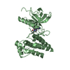
[English] 日本語
Yorodumi
- PDB-4m4x: Structure and Dimerization Properties of the Aryl Hydrocarbon Rec... -

+
Open data


ID or keywords:

Loading...

-
Basic information

Entry
Database: PDB / ID: 4m4x
TitleStructure and Dimerization Properties of the Aryl Hydrocarbon Receptor (AHR) PAS-A Domain
ComponentsAryl hydrocarbon receptor
KeywordsTRANSCRIPTION / AHR / PAS-A / Dimer / interface / transcription factor / ARNT
Function / homology
Function and homology information


circumferential growth involved in left ventricle morphogenesis / negative regulation of calcium ion transmembrane transport / cellular response to 3-methylcholanthrene / regulation of heart growth / negative regulation of T cell mediated immune response to tumor cell / cytosolic aryl hydrocarbon receptor complex / glomerulus morphogenesis / gland development / reactive oxygen species biosynthetic process / Phase I - Functionalization of compounds ...circumferential growth involved in left ventricle morphogenesis / negative regulation of calcium ion transmembrane transport / cellular response to 3-methylcholanthrene / regulation of heart growth / negative regulation of T cell mediated immune response to tumor cell / cytosolic aryl hydrocarbon receptor complex / glomerulus morphogenesis / gland development / reactive oxygen species biosynthetic process / Phase I - Functionalization of compounds / Xenobiotics / Aryl hydrocarbon receptor signalling / kidney morphogenesis / omega-hydroxylase P450 pathway / arachidonic acid omega-hydroxylase activity / positive regulation of growth rate / lymphocyte homeostasis / regulation of adaptive immune response / cellular response to 2,3,7,8-tetrachlorodibenzodioxine / Endogenous sterols / nuclear aryl hydrocarbon receptor complex / cardiac left ventricle morphogenesis / negative regulation of osteoblast proliferation / reproductive structure development / cellular response to toxic substance / prostate gland development / aryl hydrocarbon receptor complex / B-1 B cell homeostasis / post-embryonic hemopoiesis / negative regulation of DNA biosynthetic process / vasculature development / camera-type eye development / negative regulation of systemic arterial blood pressure / blood circulation / blood vessel morphogenesis / negative regulation of vasoconstriction / branching involved in blood vessel morphogenesis / immune system process / blood vessel development / E-box binding / T cell homeostasis / aryl hydrocarbon receptor binding / B cell homeostasis / positive regulation of cell size / protein localization to nucleus / toxic substance binding / blood vessel remodeling / negative regulation of osteoblast differentiation / positive regulation of RNA polymerase II transcription preinitiation complex assembly / cis-regulatory region sequence-specific DNA binding / ovarian follicle development / spleen development / xenobiotic metabolic process / B cell differentiation / liver development / circadian regulation of gene expression / Hsp90 protein binding / cell morphogenesis / response to organic cyclic compound / nuclear receptor activity / sequence-specific double-stranded DNA binding / protein-folding chaperone binding / transcription regulator complex / sequence-specific DNA binding / transcription by RNA polymerase II / transcription cis-regulatory region binding / DNA-binding transcription factor activity, RNA polymerase II-specific / response to xenobiotic stimulus / positive regulation of apoptotic process / cell cycle / protein heterodimerization activity / DNA-binding transcription factor activity / negative regulation of DNA-templated transcription / regulation of DNA-templated transcription / positive regulation of DNA-templated transcription / negative regulation of transcription by RNA polymerase II / protein homodimerization activity / DNA binding / identical protein binding / nucleus / cytoplasm / cytosol
Similarity search - Function
Aryl hydrocarbon receptor / Aryl hydrocarbon receptor/Aryl hydrocarbon receptor repressor / PAS fold-3 / PAS fold / Helix-loop-helix DNA-binding domain / PAC motif / Motif C-terminal to PAS motifs (likely to contribute to PAS structural domain) / PAS domain / Helix-loop-helix DNA-binding domain superfamily / helix loop helix domain ...Aryl hydrocarbon receptor / Aryl hydrocarbon receptor/Aryl hydrocarbon receptor repressor / PAS fold-3 / PAS fold / Helix-loop-helix DNA-binding domain / PAC motif / Motif C-terminal to PAS motifs (likely to contribute to PAS structural domain) / PAS domain / Helix-loop-helix DNA-binding domain superfamily / helix loop helix domain / Myc-type, basic helix-loop-helix (bHLH) domain / Myc-type, basic helix-loop-helix (bHLH) domain profile. / Beta-Lactamase / PAS fold / PAS fold / PAS domain / PAS repeat profile. / PAS domain / PAS domain superfamily / 2-Layer Sandwich / Alpha Beta
Similarity search - Domain/homology
Aryl hydrocarbon receptor
Similarity search - Component
Biological speciesMus musculus (house mouse)
MethodX-RAY DIFFRACTION / SYNCHROTRON / MOLECULAR REPLACEMENT / Resolution: 2.551 Å
AuthorsWu, D. / Potluri, N. / Kim, Y. / Rastinejad, F.
CitationJournal: Mol.Cell.Biol. / Year: 2013
Title: Structure and dimerization properties of the aryl hydrocarbon receptor PAS-A domain.
Authors: Wu, D. / Potluri, N. / Kim, Y. / Rastinejad, F.
History
DepositionAug 7, 2013Deposition site: RCSB / Processing site: RCSB
Revision 1.0Sep 18, 2013Provider: repository / Type: Initial release
Revision 1.1Dec 25, 2013Group: Database references
Revision 1.2Nov 15, 2017Group: Refinement description / Category: software
Revision 1.3Feb 28, 2024Group: Data collection / Database references
Category: chem_comp_atom / chem_comp_bond ...chem_comp_atom / chem_comp_bond / database_2 / struct_ref_seq_dif
Item: _database_2.pdbx_DOI / _database_2.pdbx_database_accession / _struct_ref_seq_dif.details

-
Structure visualization

Structure viewerMolecule:
MolmilJmol/JSmol

Downloads & links

-
Assembly

Deposited unit
A: Aryl hydrocarbon receptor
B: Aryl hydrocarbon receptor


Theoretical massNumber of molelcules
Total (without water)42,2592
Polymers42,2592
Non-polymers00
Water70339
1


  • Idetical with deposited unit
  • defined by author&software
TypeNameSymmetry operationNumber
identity operation1_555x,y,z1
Buried area1900 Å2
ΔGint-19 kcal/mol
Surface area12850 Å2
MethodPISA
Unit cell
Length a, b, c (Å)88.173, 88.173, 110.014
Angle α, β, γ (deg.)90.000, 90.000, 90.000
Int Tables number92
Space group name H-MP41212
Noncrystallographic symmetry (NCS)NCS domain: (Details: chain B and segid A)
NCS domain segments: (Selection details: chain 'B' and segid 'A ')

-
Components

#1: Protein Aryl hydrocarbon receptor / Ah receptor / AhR


Mass: 21129.623 Da / Num. of mol.: 2 / Fragment: PAS-A domain, UNP residues 110-267
Source method: isolated from a genetically manipulated source
Source: (gene. exp.) Mus musculus (house mouse) / Strain: C57BL / Gene: Ahr / Plasmid: pSJ2 / Production host: Escherichia coli (E. coli) / Strain (production host): BL21 / References: UniProt: P30561
#2: Water ChemComp-HOH / water


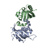
Mass: 18.015 Da / Num. of mol.: 39 / Source method: isolated from a natural source / Formula: H2O

-
Experimental details

-
Experiment

ExperimentMethod: X-RAY DIFFRACTION / Number of used crystals: 1

-
Sample preparation

CrystalDensity Matthews: 2.53 Å3/Da / Density % sol: 51.32 %
Crystal growTemperature: 277 K / Method: vapor diffusion, sitting drop / pH: 7.5
Details: 100 mM HEPES, 1.6 M Ammonium Sulfate and 2% PEG 1000, pH 7.5, VAPOR DIFFUSION, SITTING DROP, temperature 277K

-
Data collection

DiffractionMean temperature: 100 K
Diffraction sourceSource: SYNCHROTRON / Site: APS / Beamline: 19-ID / Wavelength: 0.9793 Å
DetectorType: ADSC QUANTUM 315r / Detector: CCD
RadiationScattering type: x-ray
Radiation wavelengthWavelength: 0.9793 Å / Relative weight: 1
ReflectionResolution: 2.55→50 Å / Num. obs: 14659 / % possible obs: 99.5 % / Redundancy: 9.1 % / Rmerge(I) obs: 0.07 / Χ2: 1.124 / Net I/σ(I): 11.2
Reflection shell
Resolution (Å)Redundancy (%)Rmerge(I) obsNum. unique allΧ2Diffraction-ID% possible all
2.55-2.598.10.8377350.9751100
2.59-2.648.80.7766980.991100
2.64-2.699.30.6667211.171100
2.69-2.759.40.5517161.063199.9
2.75-2.819.40.4447181.0851100
2.81-2.879.30.3837201.0411100
2.87-2.949.40.3117271.0511100
2.94-3.029.40.2397211.0541100
3.02-3.119.40.1747211.0661100
3.11-3.219.30.1517221.0571100
3.21-3.339.30.1217341.1011100
3.33-3.469.20.1017281.192199.6
3.46-3.629.30.0747281.091199.7
3.62-3.819.20.0577271.078199.7
3.81-4.059.10.057411.058199.7
4.05-4.369.20.0467371.025199.7
4.36-4.890.0487471.229199.2
4.8-5.498.90.0557501.738199.1
5.49-6.928.70.0527581.402198.2
6.92-5080.0368100.995195.5

-
Processing

Software
NameVersionClassificationNB
DENZOdata reduction
SCALEPACKdata scaling
PHENIX1.8.2_1309refinement
PDB_EXTRACT3.11data extraction
HKL-3000data collection
HKL-3000data reduction
HKL-3000data scaling
PHASERphasing
RefinementMethod to determine structure: MOLECULAR REPLACEMENT / Resolution: 2.551→33.86 Å / Occupancy max: 1 / Occupancy min: 1 / FOM work R set: 0.8256 / SU ML: 0.26 / σ(F): 1.35 / Phase error: 23.56 / Stereochemistry target values: ML
RfactorNum. reflection% reflectionSelection details
Rfree0.2446 736 5.04 %Random
Rwork0.2002 ---
obs0.2022 14617 99.49 %-
Solvent computationShrinkage radii: 0.9 Å / VDW probe radii: 1.11 Å / Solvent model: FLAT BULK SOLVENT MODEL
Displacement parametersBiso max: 160.61 Å2 / Biso mean: 60.8807 Å2 / Biso min: 25.76 Å2
Refinement stepCycle: LAST / Resolution: 2.551→33.86 Å
ProteinNucleic acidLigandSolventTotal
Num. atoms1910 0 0 39 1949
Refine LS restraints
Refine-IDTypeDev idealNumber
X-RAY DIFFRACTIONf_bond_d0.0061949
X-RAY DIFFRACTIONf_angle_d1.0982629
X-RAY DIFFRACTIONf_chiral_restr0.076291
X-RAY DIFFRACTIONf_plane_restr0.004338
X-RAY DIFFRACTIONf_dihedral_angle_d15.189696
Refine LS restraints NCSNumber: 929 / Type: POSITIONAL / Rms dev position: 12.282 Å
LS refinement shell

Refine-ID: X-RAY DIFFRACTION / Total num. of bins used: 5

Resolution (Å)Rfactor RfreeNum. reflection RfreeRfactor RworkNum. reflection RworkNum. reflection all% reflection obs (%)
2.5514-2.74830.27571540.22526982852100
2.7483-3.02480.28941450.204427382883100
3.0248-3.46210.261650.186727322897100
3.4621-4.36040.21111350.185827982933100
4.3604-33.86260.2471370.21012915305298
Refinement TLS params.

Method: refined / Refine-ID: X-RAY DIFFRACTION

IDL112)L122)L132)L222)L232)L332)S11 (Å °)S12 (Å °)S13 (Å °)S21 (Å °)S22 (Å °)S23 (Å °)S31 (Å °)S32 (Å °)S33 (Å °)T112)T122)T132)T222)T232)T332)Origin x (Å)Origin y (Å)Origin z (Å)
14.36280.83981.1413.6332-1.0666.3766-0.0739-0.02180.1139-0.18260.10770.0430.0709-0.1545-0.01410.29420.00490.02450.25850.02740.2632.18593.161712.3033
23.47181.97010.26475.1492-0.00193.3882-0.33870.58610.0224-0.55390.45820.7741-0.3011-0.2042-0.11020.4969-0.0569-0.13580.49990.14260.53958.748-10.52476.3064
Refinement TLS group
IDRefine-IDRefine TLS-IDSelection detailsAuth asym-IDAuth seq-ID
1X-RAY DIFFRACTION1(chain A and resseq -10:9999)A107 - 266
2X-RAY DIFFRACTION2(chain B and resseq -10:9999)B108 - 266

+
About Yorodumi

-
News

-
Feb 9, 2022. New format data for meta-information of EMDB entries

New format data for meta-information of EMDB entries

  • Version 3 of the EMDB header file is now the official format.
  • The previous official version 1.9 will be removed from the archive.

Related info.:EMDB header

External links:wwPDB to switch to version 3 of the EMDB data model

-
Aug 12, 2020. Covid-19 info

Covid-19 info

URL: https://pdbjlvh1.pdbj.org/emnavi/covid19.php

New page: Covid-19 featured information page in EM Navigator.

Related info.:Covid-19 info / Mar 5, 2020. Novel coronavirus structure data

+
Mar 5, 2020. Novel coronavirus structure data

Novel coronavirus structure data

Related info.:Yorodumi Speices / Aug 12, 2020. Covid-19 info

External links:COVID-19 featured content - PDBj / Molecule of the Month (242):Coronavirus Proteases

+
Jan 31, 2019. EMDB accession codes are about to change! (news from PDBe EMDB page)

EMDB accession codes are about to change! (news from PDBe EMDB page)

  • The allocation of 4 digits for EMDB accession codes will soon come to an end. Whilst these codes will remain in use, new EMDB accession codes will include an additional digit and will expand incrementally as the available range of codes is exhausted. The current 4-digit format prefixed with “EMD-” (i.e. EMD-XXXX) will advance to a 5-digit format (i.e. EMD-XXXXX), and so on. It is currently estimated that the 4-digit codes will be depleted around Spring 2019, at which point the 5-digit format will come into force.
  • The EM Navigator/Yorodumi systems omit the EMD- prefix.

Related info.:Q: What is EMD? / ID/Accession-code notation in Yorodumi/EM Navigator

External links:EMDB Accession Codes are Changing Soon! / Contact to PDBj

+
Jul 12, 2017. Major update of PDB

Major update of PDB

  • wwPDB released updated PDB data conforming to the new PDBx/mmCIF dictionary.
  • This is a major update changing the version number from 4 to 5, and with Remediation, in which all the entries are updated.
  • In this update, many items about electron microscopy experimental information are reorganized (e.g. em_software).
  • Now, EM Navigator and Yorodumi are based on the updated data.

External links:wwPDB Remediation / Enriched Model Files Conforming to OneDep Data Standards Now Available in the PDB FTP Archive

-
Yorodumi

Thousand views of thousand structures

  • Yorodumi is a browser for structure data from EMDB, PDB, SASBDB, etc.
  • This page is also the successor to EM Navigator detail page, and also detail information page/front-end page for Omokage search.
  • The word "yorodu" (or yorozu) is an old Japanese word meaning "ten thousand". "mi" (miru) is to see.

Related info.:EMDB / PDB / SASBDB / Comparison of 3 databanks / Yorodumi Search / Aug 31, 2016. New EM Navigator & Yorodumi / Yorodumi Papers / Jmol/JSmol / Function and homology information / Changes in new EM Navigator and Yorodumi

Read more