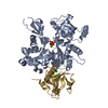
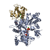
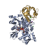
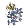
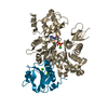
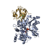
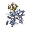
Journal: PLoS Pathog / Year: 2014 Title: Structural differences explain diverse functions of Plasmodium actins. Authors: Juha Vahokoski / Saligram Prabhakar Bhargav / Ambroise Desfosses / Maria Andreadaki / Esa-Pekka Kumpula / Silvia Muñico Martinez / Alexander Ignatev / Simone Lepper / Friedrich Frischknecht ...Authors: Juha Vahokoski / Saligram Prabhakar Bhargav / Ambroise Desfosses / Maria Andreadaki / Esa-Pekka Kumpula / Silvia Muñico Martinez / Alexander Ignatev / Simone Lepper / Friedrich Frischknecht / Inga Sidén-Kiamos / Carsten Sachse / Inari Kursula / Abstract: Actins are highly conserved proteins and key players in central processes in all eukaryotic cells. The two actins of the malaria parasite are among the most divergent eukaryotic actins and also ...Actins are highly conserved proteins and key players in central processes in all eukaryotic cells. The two actins of the malaria parasite are among the most divergent eukaryotic actins and also differ from each other more than isoforms in any other species. Microfilaments have not been directly observed in Plasmodium and are presumed to be short and highly dynamic. We show that actin I cannot complement actin II in male gametogenesis, suggesting critical structural differences. Cryo-EM reveals that Plasmodium actin I has a unique filament structure, whereas actin II filaments resemble canonical F-actin. Both Plasmodium actins hydrolyze ATP more efficiently than α-actin, and unlike any other actin, both parasite actins rapidly form short oligomers induced by ADP. Crystal structures of both isoforms pinpoint several structural changes in the monomers causing the unique polymerization properties. Inserting the canonical D-loop to Plasmodium actin I leads to the formation of long filaments in vitro. In vivo, this chimera restores gametogenesis in parasites lacking actin II, suggesting that stable filaments are required for exflagellation. Together, these data underline the divergence of eukaryotic actins and demonstrate how structural differences in the monomers translate into filaments with different properties, implying that even eukaryotic actins have faced different evolutionary pressures and followed different paths for developing their polymerization properties.
In the structure databanks used in Yorodumi, some data are registered as the other names, "COVID-19 virus" and "2019-nCoV". Here are the details of the virus and the list of structure data.
Jan 31, 2019. EMDB accession codes are about to change! (news from PDBe EMDB page)
EMDB accession codes are about to change! (news from PDBe EMDB page)
The allocation of 4 digits for EMDB accession codes will soon come to an end. Whilst these codes will remain in use, new EMDB accession codes will include an additional digit and will expand incrementally as the available range of codes is exhausted. The current 4-digit format prefixed with “EMD-” (i.e. EMD-XXXX) will advance to a 5-digit format (i.e. EMD-XXXXX), and so on. It is currently estimated that the 4-digit codes will be depleted around Spring 2019, at which point the 5-digit format will come into force.
The EM Navigator/Yorodumi systems omit the EMD- prefix.
Related info.:Q: What is EMD? / ID/Accession-code notation in Yorodumi/EM Navigator
Yorodumi is a browser for structure data from EMDB, PDB, SASBDB, etc.
This page is also the successor to EM Navigator detail page, and also detail information page/front-end page for Omokage search.
The word "yorodu" (or yorozu) is an old Japanese word meaning "ten thousand". "mi" (miru) is to see.
Related info.:EMDB / PDB / SASBDB / Comparison of 3 databanks / Yorodumi Search / Aug 31, 2016. New EM Navigator & Yorodumi / Yorodumi Papers / Jmol/JSmol / Function and homology information / Changes in new EM Navigator and Yorodumi
 Movie
Movie Controller


 Open data
 Basic information
 Components
 Keywords
 Function and homology information

 X-RAY DIFFRACTION /
  Authors
 Citation


 Structure visualization
 Downloads & links
 Other downloads
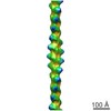


 PDBj
PDBj


 Assembly

 SPODOPTERA FRUGIPERDA (fall armyworm) / Strain (production host): SF21

 Mass: 507.181 Da / Num. of mol.: 1 / Source method: obtained synthetically / Formula: C10H16N5O13P3 / Comment: ATP, energy-carrying molecule*YM
 Mass: 40.078 Da / Num. of mol.: 3 / Source method: obtained synthetically / Formula: Ca
 Mass: 18.015 Da / Num. of mol.: 774 / Source method: isolated from a natural source / Formula: H2O
 Sample preparation
 Processing