 Movie
Movie Controller

[English] 日本語
 Yorodumi
- PDB-6i4d: Crystal Structure of Plasmodium falciparum actin I in the Mg-K-AT... -
+ Open data

- Basic information
| Entry | Database: PDB / ID: 6i4d | ||||||||||||
|---|---|---|---|---|---|---|---|---|---|---|---|---|---|
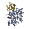
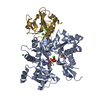
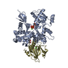
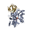
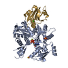
| Title | Crystal Structure of Plasmodium falciparum actin I in the Mg-K-ATP/ADP state | ||||||||||||
 Components Components |
| ||||||||||||
 Keywords Keywords | CONTRACTILE PROTEIN / hydrolase / filamentous / glideosome / cytoskeleton | ||||||||||||
| Function / homology |  Function and homology information Function and homology informationglial filament / plastid inheritance / schizogony / basal ectoplasmic specialization / apical ectoplasmic specialization / symbiont-mediated actin polymerization-dependent cell-to-cell migration in host / Platelet degranulation / Caspase-mediated cleavage of cytoskeletal proteins / regulation of podosome assembly / striated muscle atrophy ...glial filament / plastid inheritance / schizogony / basal ectoplasmic specialization / apical ectoplasmic specialization / symbiont-mediated actin polymerization-dependent cell-to-cell migration in host / Platelet degranulation / Caspase-mediated cleavage of cytoskeletal proteins / regulation of podosome assembly / striated muscle atrophy / regulation of establishment of T cell polarity / regulation of plasma membrane raft polarization / regulation of receptor clustering / positive regulation of keratinocyte apoptotic process / renal protein absorption / positive regulation of protein processing in phagocytic vesicle / phosphatidylinositol 3-kinase catalytic subunit binding / positive regulation of actin nucleation / actin cap / myosin II binding / host-mediated suppression of symbiont invasion / cell projection assembly / entry into host cell by a symbiont-containing vacuole / actin filament severing / barbed-end actin filament capping / actin polymerization or depolymerization / actin filament depolymerization / actin filament capping / positive regulation of p38MAPK cascade / podosome / relaxation of cardiac muscle / phagocytosis, engulfment / cardiac muscle cell contraction / hepatocyte apoptotic process / cortical actin cytoskeleton / positive regulation of cardiac muscle hypertrophy / cellular response to cadmium ion / sarcoplasm / cilium assembly / phagocytic vesicle / ruffle / response to muscle stretch / vesicle-mediated transport / actin filament polymerization / Neutrophil degranulation / cytoskeleton organization / phosphatidylinositol-4,5-bisphosphate binding / actin filament organization / central nervous system development / actin filament / cellular response to type II interferon / protein destabilization / structural constituent of cytoskeleton / Hydrolases; Acting on acid anhydrides; Acting on acid anhydrides to facilitate cellular and subcellular movement / actin filament binding / myelin sheath / lamellipodium / actin cytoskeleton / actin cytoskeleton organization / actin binding / amyloid fibril formation / calcium ion binding / positive regulation of gene expression / perinuclear region of cytoplasm / ATP hydrolysis activity / protein-containing complex / : / extracellular region / ATP binding / nucleus / plasma membrane / cytosol / cytoplasm Similarity search - Function | ||||||||||||
| Biological species |   | ||||||||||||
| Method |  X-RAY DIFFRACTION / SYNCHROTRON / MOLECULAR REPLACEMENT / Resolution: 1.24 Å X-RAY DIFFRACTION / SYNCHROTRON / MOLECULAR REPLACEMENT / Resolution: 1.24 Å | ||||||||||||
 Authors Authors | Kumpula, E.-P. / Lopez, A.J. / Tajedin, L. / Han, H. / Kursula, I. | ||||||||||||
| Funding support |  Finland, Finland,  Norway, 3items Norway, 3items
| ||||||||||||
 Citation Citation | Journal: Plos Biol. / Year: 2019 Title: Atomic view into Plasmodium actin polymerization, ATP hydrolysis, and fragmentation. Authors: Kumpula, E.P. / Lopez, A.J. / Tajedin, L. / Han, H. / Kursula, I. | ||||||||||||
| History |
|
- Structure visualization
| Structure viewer | Molecule: MolmilJmol/JSmol |
|---|
- Downloads & links
-Download
| PDBx/mmCIF format | 6i4d.cif.gz | 443.4 KB | Display | PDBx/mmCIF format |
|---|---|---|---|---|
| PDB format | pdb6i4d.ent.gz | 305.7 KB | Display | PDB format |
| PDBx/mmJSON format | 6i4d.json.gz | Tree view | PDBx/mmJSON format | |
| Others |  Other downloads Other downloads |
-Validation report
| Arichive directory | https://data.pdbj.org/pub/pdb/validation_reports/i4/6i4dftp://data.pdbj.org/pub/pdb/validation_reports/i4/6i4d | HTTPS FTP |
|---|
-Related structure data
| Related structure data |  6i4eC  6i4fC  6i4gC  6i4hC 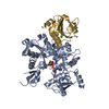 6i4iC  6i4jC  6i4kC  6i4lC  6i4mC 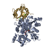 4cbuS S: Starting model for refinement C: citing same article ( |
|---|---|
| Similar structure data |

-Links
 PDBj
PDBj


- Assembly
| Deposited unit | 
| ||||||||||||
|---|---|---|---|---|---|---|---|---|---|---|---|---|---|
| 1 |
| ||||||||||||
| Unit cell |
| ||||||||||||
| Components on special symmetry positions |
|
-Components
-Protein , 2 types, 2 molecules AG
| #1: Protein | Mass: 42047.676 Da / Num. of mol.: 1 Source method: isolated from a genetically manipulated source Source: (gene. exp.) Gene: PFL2215w / Production host:   Spodoptera frugiperda (fall armyworm) / References: UniProt: Q8I4X0 Spodoptera frugiperda (fall armyworm) / References: UniProt: Q8I4X0 |
|---|---|
| #2: Protein | Mass: 14239.979 Da / Num. of mol.: 1 Source method: isolated from a genetically manipulated source Source: (gene. exp.)  |
-Non-polymers , 9 types, 699 molecules 


| #3: Chemical | ChemComp-ATP /  Mass: 507.181 Da / Num. of mol.: 1 / Source method: obtained synthetically / Formula: C10H16N5O13P3 / Feature type: SUBJECT OF INVESTIGATION / Comment: ATP, energy-carrying molecule*YM Mass: 507.181 Da / Num. of mol.: 1 / Source method: obtained synthetically / Formula: C10H16N5O13P3 / Feature type: SUBJECT OF INVESTIGATION / Comment: ATP, energy-carrying molecule*YM | ||||||||
|---|---|---|---|---|---|---|---|---|---|
| #4: Chemical | ChemComp-ADP /  Mass: 427.201 Da / Num. of mol.: 1 / Source method: obtained synthetically / Formula: C10H15N5O10P2 / Feature type: SUBJECT OF INVESTIGATION / Comment: ADP, energy-carrying molecule*YM Mass: 427.201 Da / Num. of mol.: 1 / Source method: obtained synthetically / Formula: C10H15N5O10P2 / Feature type: SUBJECT OF INVESTIGATION / Comment: ADP, energy-carrying molecule*YM | ||||||||
| #5: Chemical | ChemComp-MG /  Mass: 24.305 Da / Num. of mol.: 1 / Source method: obtained synthetically / Formula: Mg / Feature type: SUBJECT OF INVESTIGATION Mass: 24.305 Da / Num. of mol.: 1 / Source method: obtained synthetically / Formula: Mg / Feature type: SUBJECT OF INVESTIGATION | ||||||||
| #6: Chemical | ChemComp-K /  Mass: 39.098 Da / Num. of mol.: 1 / Source method: obtained synthetically / Formula: K / Feature type: SUBJECT OF INVESTIGATION Mass: 39.098 Da / Num. of mol.: 1 / Source method: obtained synthetically / Formula: K / Feature type: SUBJECT OF INVESTIGATION | ||||||||
| #7: Chemical |  Mass: 58.082 Da / Num. of mol.: 2 / Source method: obtained synthetically / Formula: CNS Mass: 58.082 Da / Num. of mol.: 2 / Source method: obtained synthetically / Formula: CNS#8: Chemical | ChemComp-BTB / |  Mass: 209.240 Da / Num. of mol.: 1 / Source method: obtained synthetically / Formula: C8H19NO5 / Comment: pH buffer*YM Mass: 209.240 Da / Num. of mol.: 1 / Source method: obtained synthetically / Formula: C8H19NO5 / Comment: pH buffer*YM#9: Chemical | ChemComp-CL / |  Mass: 35.453 Da / Num. of mol.: 1 / Source method: obtained synthetically / Formula: Cl Mass: 35.453 Da / Num. of mol.: 1 / Source method: obtained synthetically / Formula: Cl#10: Chemical |  Mass: 40.078 Da / Num. of mol.: 2 / Source method: obtained synthetically / Formula: Ca Mass: 40.078 Da / Num. of mol.: 2 / Source method: obtained synthetically / Formula: Ca#11: Water | ChemComp-HOH / | Mass: 18.015 Da / Num. of mol.: 689 / Source method: isolated from a natural source / Formula: H2O |
-Experimental details
-Experiment
| Experiment | Method: X-RAY DIFFRACTION / Number of used crystals: 1 |
|---|
- Sample preparation
| Crystal | Density Matthews: 2.39 Å3/Da / Density % sol: 48.59 % |
|---|---|
| Crystal grow | Temperature: 298 K / Method: vapor diffusion, sitting drop / pH: 6 Details: 23%(w/v) PEG3350, 0.1 M BIS-TRIS pH 6.0, 0.2M K-SCN; 20% PEG400 used for cryoprotection |
-Data collection
| Diffraction | Mean temperature: 100 K / Serial crystal experiment: N |
|---|---|
| Diffraction source | Source: SYNCHROTRON / Site: PETRA III, EMBL c/o DESY  / Beamline: P13 (MX1) / Wavelength: 1.031 Å / Beamline: P13 (MX1) / Wavelength: 1.031 Å |
| Detector | Type: DECTRIS PILATUS 6M / Detector: PIXEL / Date: Sep 19, 2013 |
| Radiation | Protocol: SINGLE WAVELENGTH / Monochromatic (M) / Laue (L): M / Scattering type: x-ray |
| Radiation wavelength | Wavelength: 1.031 Å / Relative weight: 1 |
| Reflection | Resolution: 1.24→59.93 Å / Num. obs: 152407 / % possible obs: 98.8 % / Redundancy: 7 % / Biso Wilson estimate: 12.4 Å2 / CC1/2: 1 / Rmerge(I) obs: 0.062 / Net I/σ(I): 17.5 |
| Reflection shell | Resolution: 1.24→1.28 Å / Redundancy: 4.9 % / Rmerge(I) obs: 1.071 / Mean I/σ(I) obs: 1.3 / Num. unique obs: 13428 / CC1/2: 0.509 / Rpim(I) all: 0.5109 / Rrim(I) all: 1.193 / % possible all: 88.2 |
- Processing
| Software |
| |||||||||||||||||||||||||||||||||||||||||||||||||||||||||||||||||||||||||||||||||||||||||||||||||||||||||
|---|---|---|---|---|---|---|---|---|---|---|---|---|---|---|---|---|---|---|---|---|---|---|---|---|---|---|---|---|---|---|---|---|---|---|---|---|---|---|---|---|---|---|---|---|---|---|---|---|---|---|---|---|---|---|---|---|---|---|---|---|---|---|---|---|---|---|---|---|---|---|---|---|---|---|---|---|---|---|---|---|---|---|---|---|---|---|---|---|---|---|---|---|---|---|---|---|---|---|---|---|---|---|---|---|---|---|
| Refinement | Method to determine structure: MOLECULAR REPLACEMENT Starting model: 4CBU Resolution: 1.24→59.93 Å / SU ML: 0.1323 / Cross valid method: FREE R-VALUE / σ(F): 1.4 / Phase error: 14.6308
| |||||||||||||||||||||||||||||||||||||||||||||||||||||||||||||||||||||||||||||||||||||||||||||||||||||||||
| Solvent computation | Shrinkage radii: 0.9 Å / VDW probe radii: 1.11 Å | |||||||||||||||||||||||||||||||||||||||||||||||||||||||||||||||||||||||||||||||||||||||||||||||||||||||||
| Displacement parameters | Biso mean: 18.76 Å2 | |||||||||||||||||||||||||||||||||||||||||||||||||||||||||||||||||||||||||||||||||||||||||||||||||||||||||
| Refinement step | Cycle: LAST / Resolution: 1.24→59.93 Å
| |||||||||||||||||||||||||||||||||||||||||||||||||||||||||||||||||||||||||||||||||||||||||||||||||||||||||
| Refine LS restraints |
| |||||||||||||||||||||||||||||||||||||||||||||||||||||||||||||||||||||||||||||||||||||||||||||||||||||||||
| LS refinement shell |
|
