 Movie
Movie Controller

[English] 日本語
 Yorodumi
- PDB-4c3p: Structure of dephosphorylated Aurora A (122-403) bound to TPX2 an... -
+ Open data

- Basic information
| Entry | Database: PDB / ID: 4c3p | ||||||
|---|---|---|---|---|---|---|---|
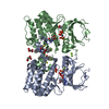
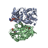
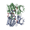
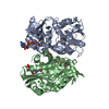
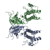
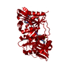
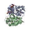
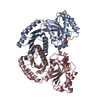
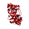
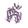
| Title | Structure of dephosphorylated Aurora A (122-403) bound to TPX2 and AMPPCP | ||||||
 Components Components |
| ||||||
 Keywords Keywords | TRANSFERASE / ACTIVATION / ALLOSTERY / CELL CYCLE / CANCER | ||||||
| Function / homology |  Function and homology information Function and homology informationactivation of protein kinase activity / Interaction between PHLDA1 and AURKA / regulation of centrosome cycle / axon hillock / negative regulation of microtubule depolymerization / spindle assembly involved in female meiosis I / cilium disassembly / spindle pole centrosome / importin-alpha family protein binding / microtubule nucleation ...activation of protein kinase activity / Interaction between PHLDA1 and AURKA / regulation of centrosome cycle / axon hillock / negative regulation of microtubule depolymerization / spindle assembly involved in female meiosis I / cilium disassembly / spindle pole centrosome / importin-alpha family protein binding / microtubule nucleation / chromosome passenger complex / histone H3S10 kinase activity / positive regulation of oocyte maturation / mitotic centrosome separation / pronucleus / germinal vesicle / protein localization to centrosome / meiotic spindle / anterior/posterior axis specification / neuron projection extension / spindle organization / centrosome localization / positive regulation of mitochondrial fission / mitotic spindle pole / spindle midzone / SUMOylation of DNA replication proteins / mitotic spindle assembly / negative regulation of protein binding / intercellular bridge / regulation of G2/M transition of mitotic cell cycle / liver regeneration / protein serine/threonine/tyrosine kinase activity / centriole / positive regulation of mitotic nuclear division / positive regulation of mitotic cell cycle / regulation of mitotic spindle organization / TP53 Regulates Transcription of Genes Involved in G2 Cell Cycle Arrest / molecular function activator activity / regulation of signal transduction by p53 class mediator / AURKA Activation by TPX2 / protein serine/threonine kinase activator activity / mitotic spindle organization / regulation of cytokinesis / FBXL7 down-regulates AURKA during mitotic entry and in early mitosis / APC/C:Cdh1 mediated degradation of Cdc20 and other APC/C:Cdh1 targeted proteins in late mitosis/early G1 / peptidyl-serine phosphorylation / regulation of protein stability / kinetochore / response to wounding / G2/M transition of mitotic cell cycle / spindle / spindle pole / mitotic spindle / Regulation of PLK1 Activity at G2/M Transition / positive regulation of proteasomal ubiquitin-dependent protein catabolic process / mitotic cell cycle / protein autophosphorylation / microtubule cytoskeleton / midbody / basolateral plasma membrane / molecular adaptor activity / Regulation of TP53 Activity through Phosphorylation / proteasome-mediated ubiquitin-dependent protein catabolic process / microtubule / protein phosphorylation / non-specific serine/threonine protein kinase / protein kinase activity / postsynaptic density / ciliary basal body / protein heterodimerization activity / negative regulation of gene expression / cell division / protein serine kinase activity / protein serine/threonine kinase activity / apoptotic process / ubiquitin protein ligase binding / centrosome / protein kinase binding / negative regulation of apoptotic process / perinuclear region of cytoplasm / glutamatergic synapse / nucleoplasm / ATP binding / nucleus / cytosol Similarity search - Function | ||||||
| Biological species |  HOMO SAPIENS (human) HOMO SAPIENS (human) | ||||||
| Method |  X-RAY DIFFRACTION / SYNCHROTRON / MOLECULAR REPLACEMENT / Resolution: 2.69 Å X-RAY DIFFRACTION / SYNCHROTRON / MOLECULAR REPLACEMENT / Resolution: 2.69 Å | ||||||
 Authors Authors | Zorba, A. / Kutter, S. / Kern, D. | ||||||
 Citation Citation | Journal: Elife / Year: 2014 Title: Molecular Mechanism of Aurora a Kinase Autophosphorylation and its Allosteric Activation by Tpx2. Authors: Zorba, A. / Buosi, V. / Kutter, S. / Kern, N. / Pontiggia, F. / Cho, Y.J. / Kern, D. | ||||||
| History |
|
- Structure visualization
| Structure viewer | Molecule: MolmilJmol/JSmol |
|---|
- Downloads & links
-Download
| PDBx/mmCIF format | 4c3p.cif.gz | 129.7 KB | Display | PDBx/mmCIF format |
|---|---|---|---|---|
| PDB format | pdb4c3p.ent.gz | 99.8 KB | Display | PDB format |
| PDBx/mmJSON format | 4c3p.json.gz | Tree view | PDBx/mmJSON format | |
| Others |  Other downloads Other downloads |
-Validation report
| Summary document | 4c3p_validation.pdf.gz | 1 MB | Display | wwPDB validaton report |
|---|---|---|---|---|
| Full document | 4c3p_full_validation.pdf.gz | 1 MB | Display | |
| Data in XML | 4c3p_validation.xml.gz | 22.2 KB | Display | |
| Data in CIF | 4c3p_validation.cif.gz | 29.7 KB | Display | |
| Arichive directory | https://data.pdbj.org/pub/pdb/validation_reports/c3/4c3pftp://data.pdbj.org/pub/pdb/validation_reports/c3/4c3p | HTTPS FTP |
-Related structure data
| Related structure data | 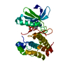 4c3rC 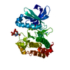 1ol7S C: citing same article ( S: Starting model for refinement |
|---|---|
| Similar structure data |
-Links
 PDBj
PDBj


- Assembly
| Deposited unit | 
| ||||||||
|---|---|---|---|---|---|---|---|---|---|
| 1 |
| ||||||||
| Unit cell |
|
-Components
| #1: Protein | Mass: 32689.459 Da / Num. of mol.: 2 / Fragment: KINASE DOMAIN, RESIDUES 121-403 Source method: isolated from a genetically manipulated source Source: (gene. exp.) HOMO SAPIENS (human) / Plasmid: PET28A / Production host:  References: UniProt: O14965, non-specific serine/threonine protein kinase #2: Protein/peptide | Mass: 4927.110 Da / Num. of mol.: 2 / Fragment: AURORA A KINASE BINDING DOMAIN, RESIDUES 1-43 Source method: isolated from a genetically manipulated source Source: (gene. exp.) HOMO SAPIENS (human) / Plasmid: PET28A / Production host: #3: Chemical |   Mass: 505.208 Da / Num. of mol.: 2 / Source method: obtained synthetically / Formula: C11H18N5O12P3 / Comment: AMP-PCP, energy-carrying molecule analogue*YM Mass: 505.208 Da / Num. of mol.: 2 / Source method: obtained synthetically / Formula: C11H18N5O12P3 / Comment: AMP-PCP, energy-carrying molecule analogue*YM#4: Chemical | ChemComp-SO4 / |   Mass: 96.063 Da / Num. of mol.: 1 / Source method: obtained synthetically / Formula: SO4 Mass: 96.063 Da / Num. of mol.: 1 / Source method: obtained synthetically / Formula: SO4#5: Water | ChemComp-HOH / |  Mass: 18.015 Da / Num. of mol.: 21 / Source method: isolated from a natural source / Formula: H2O Mass: 18.015 Da / Num. of mol.: 21 / Source method: isolated from a natural source / Formula: H2O |
|---|
-Experimental details
-Experiment
| Experiment | Method: X-RAY DIFFRACTION / Number of used crystals: 1 |
|---|
- Sample preparation
| Crystal | Density Matthews: 2.57 Å3/Da / Density % sol: 52.23 % / Description: NONE |
|---|---|
| Crystal grow | Temperature: 291 K / Method: vapor diffusion, hanging drop / pH: 5.5 Details: CRYSTALS OF DEPHOSPHORYLATED A(122-403) IN COMPLEX WITH AMPPCP AND TPX2(1-43) WERE GROWN AT 18C BY VAPOR DIFFUSION AND THE HANGING DROP OBTAINED BY COMBINING 300UM DEP A(122-403) WITH 1.5MM ...Details: CRYSTALS OF DEPHOSPHORYLATED A(122-403) IN COMPLEX WITH AMPPCP AND TPX2(1-43) WERE GROWN AT 18C BY VAPOR DIFFUSION AND THE HANGING DROP OBTAINED BY COMBINING 300UM DEP A(122-403) WITH 1.5MM AMPPCP AND 300UM TPX2(1-43) WITH 0.2M LITHIUM SULFATE MONOHYDRATE, 0.1M BISTRIS PH5.5, 25% PEG3350. |
-Data collection
| Diffraction | Mean temperature: 100 K |
|---|---|
| Diffraction source | Source: SYNCHROTRON / Site: ALS  / Beamline: 8.2.2 / Wavelength: 1.21433 / Beamline: 8.2.2 / Wavelength: 1.21433 |
| Detector | Type: ADSC QUANTUM 315 / Detector: CCD / Date: Dec 17, 2012 |
| Radiation | Monochromator: DOUBLE CRYSTAL, SI(111) / Protocol: SINGLE WAVELENGTH / Monochromatic (M) / Laue (L): M / Scattering type: x-ray |
| Radiation wavelength | Wavelength: 1.21433 Å / Relative weight: 1 |
| Reflection | Resolution: 2.69→47.5 Å / Num. obs: 19139 / % possible obs: 100 % / Observed criterion σ(I): 2.3 / Redundancy: 6.9 % / Rmerge(I) obs: 0.26 / Net I/σ(I): 8 |
| Reflection shell | Resolution: 2.69→2.76 Å / Redundancy: 7.2 % / Mean I/σ(I) obs: 2.3 / % possible all: 100 |
- Processing
| Software |
| ||||||||||||||||||||||||||||||||||||||||||||||||||||||||||||||||||||||||||||||||||||||||||||||||||||||||||||||||||||||||||||||||||||||||||||||||||||||||||||||||||||||||||||||||||||||
|---|---|---|---|---|---|---|---|---|---|---|---|---|---|---|---|---|---|---|---|---|---|---|---|---|---|---|---|---|---|---|---|---|---|---|---|---|---|---|---|---|---|---|---|---|---|---|---|---|---|---|---|---|---|---|---|---|---|---|---|---|---|---|---|---|---|---|---|---|---|---|---|---|---|---|---|---|---|---|---|---|---|---|---|---|---|---|---|---|---|---|---|---|---|---|---|---|---|---|---|---|---|---|---|---|---|---|---|---|---|---|---|---|---|---|---|---|---|---|---|---|---|---|---|---|---|---|---|---|---|---|---|---|---|---|---|---|---|---|---|---|---|---|---|---|---|---|---|---|---|---|---|---|---|---|---|---|---|---|---|---|---|---|---|---|---|---|---|---|---|---|---|---|---|---|---|---|---|---|---|---|---|---|---|
| Refinement | Method to determine structure: MOLECULAR REPLACEMENT Starting model: PDB ENTRY 1OL7 Resolution: 2.69→47.48 Å / Cor.coef. Fo:Fc: 0.936 / Cor.coef. Fo:Fc free: 0.882 / SU B: 14.978 / SU ML: 0.307 / Cross valid method: THROUGHOUT / ESU R Free: 0.402 / Stereochemistry target values: MAXIMUM LIKELIHOOD Details: HYDROGENS HAVE BEEN ADDED IN THE RIDING POSITIONS. U VALUES REFINED INDIVIDUALLY
| ||||||||||||||||||||||||||||||||||||||||||||||||||||||||||||||||||||||||||||||||||||||||||||||||||||||||||||||||||||||||||||||||||||||||||||||||||||||||||||||||||||||||||||||||||||||
| Solvent computation | Ion probe radii: 0.8 Å / Shrinkage radii: 0.8 Å / VDW probe radii: 1.2 Å / Solvent model: MASK | ||||||||||||||||||||||||||||||||||||||||||||||||||||||||||||||||||||||||||||||||||||||||||||||||||||||||||||||||||||||||||||||||||||||||||||||||||||||||||||||||||||||||||||||||||||||
| Displacement parameters | Biso mean: 53.94 Å2
| ||||||||||||||||||||||||||||||||||||||||||||||||||||||||||||||||||||||||||||||||||||||||||||||||||||||||||||||||||||||||||||||||||||||||||||||||||||||||||||||||||||||||||||||||||||||
| Refinement step | Cycle: LAST / Resolution: 2.69→47.48 Å
| ||||||||||||||||||||||||||||||||||||||||||||||||||||||||||||||||||||||||||||||||||||||||||||||||||||||||||||||||||||||||||||||||||||||||||||||||||||||||||||||||||||||||||||||||||||||
| Refine LS restraints |
|