 Movie
Movie Controller


+ Open data

- Basic information
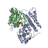
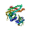
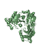
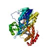
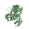
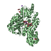
| Entry | Database: PDB / ID: 4bqz | ||||||
|---|---|---|---|---|---|---|---|
| Title | Rat NTPDase2 in complex with Mg GMPPNP | ||||||
 Components Components | ECTONUCLEOSIDE TRIPHOSPHATE DIPHOSPHOHYDROLASE 2 | ||||||
 Keywords Keywords | HYDROLASE / APYRASE / ATPASE / ADPASE / CD39 / PURINERGIC SIGNALLING / DOMAIN ROTATION / TRANSITION STATE / NTPDASE | ||||||
| Function / homology |  Function and homology information Function and homology informationribonucleoside triphosphate catabolic process / purine ribonucleoside diphosphate catabolic process / Phosphate bond hydrolysis by NTPDase proteins / apyrase activity / apyrase / UDP phosphatase activity / GDP phosphatase activity / ADP phosphatase activity / nucleoside diphosphate catabolic process / cellular response to interferon-alpha ...ribonucleoside triphosphate catabolic process / purine ribonucleoside diphosphate catabolic process / Phosphate bond hydrolysis by NTPDase proteins / apyrase activity / apyrase / UDP phosphatase activity / GDP phosphatase activity / ADP phosphatase activity / nucleoside diphosphate catabolic process / cellular response to interferon-alpha / nucleoside diphosphate phosphatase activity / cell projection membrane / response to auditory stimulus / cellular response to interleukin-6 / basement membrane / platelet activation / cellular response to tumor necrosis factor / ribonucleoside triphosphate phosphatase activity / cellular response to lipopolysaccharide / cell body / G protein-coupled receptor signaling pathway / cell surface / ATP hydrolysis activity / ATP binding / membrane / identical protein binding / plasma membrane Similarity search - Function | ||||||
| Biological species |  | ||||||
| Method |  X-RAY DIFFRACTION / SYNCHROTRON / OTHER / Resolution: 2.05 Å X-RAY DIFFRACTION / SYNCHROTRON / OTHER / Resolution: 2.05 Å | ||||||
 Authors Authors | Zebisch, M. / Schaefer, P. / Lauble, P. / Straeter, N. | ||||||
 Citation Citation | Journal: Structure / Year: 2013 Title: Crystallographic Snapshots Along the Reaction Pathway of Nucleoside Triphosphate Diphosphohydrolases Authors: Zebisch, M. / Krauss, M. / Schaefer, P. / Lauble, P. / Straeter, N. | ||||||
| History |
|
- Structure visualization
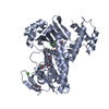
| Structure viewer | Molecule: MolmilJmol/JSmol |
|---|
- Downloads & links
-Download
| PDBx/mmCIF format | 4bqz.cif.gz | 182.2 KB | Display | PDBx/mmCIF format |
|---|---|---|---|---|
| PDB format | pdb4bqz.ent.gz | 144.4 KB | Display | PDB format |
| PDBx/mmJSON format | 4bqz.json.gz | Tree view | PDBx/mmJSON format | |
| Others |  Other downloads Other downloads |
-Validation report
| Arichive directory | https://data.pdbj.org/pub/pdb/validation_reports/bq/4bqzftp://data.pdbj.org/pub/pdb/validation_reports/bq/4bqz | HTTPS FTP |
|---|
-Related structure data
| Related structure data |  4br0C  4br2C 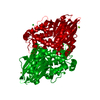 4br4C  4br5C  4br7C 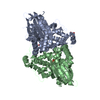 4br9C 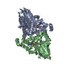 4braC 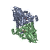 4brcC 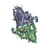 4brdC 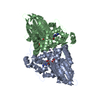 4breC 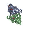 4brfC 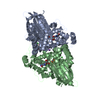 4brgC 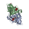 4brhC 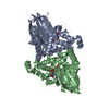 4briC  4brkC 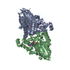 4brlC 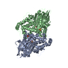 4brmC  4brnC 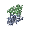 4broC  4brpC  4brqC C: citing same article ( |
|---|---|
| Similar structure data |


-Links
 PDBj
PDBj


- Assembly
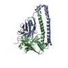
| Deposited unit | 
| ||||||||
|---|---|---|---|---|---|---|---|---|---|
| 1 |
| ||||||||
| Unit cell |
|
-Components
| #1: Protein | Mass: 50699.027 Da / Num. of mol.: 1 / Fragment: ECTODOMAIN, RESIDUES 28-462 Source method: isolated from a genetically manipulated source Source: (gene. exp.)  | ||||
|---|---|---|---|---|---|
| #2: Chemical | ChemComp-GNP /   Mass: 522.196 Da / Num. of mol.: 1 / Source method: obtained synthetically / Formula: C10H17N6O13P3 Mass: 522.196 Da / Num. of mol.: 1 / Source method: obtained synthetically / Formula: C10H17N6O13P3Comment: GppNHp, GMPPNP, energy-carrying molecule analogue*YM | ||||
| #3: Chemical | ChemComp-MG /   Mass: 24.305 Da / Num. of mol.: 1 / Source method: obtained synthetically / Formula: Mg Mass: 24.305 Da / Num. of mol.: 1 / Source method: obtained synthetically / Formula: Mg | ||||
| #4: Chemical |   Mass: 92.094 Da / Num. of mol.: 3 / Source method: obtained synthetically / Formula: C3H8O3 Mass: 92.094 Da / Num. of mol.: 3 / Source method: obtained synthetically / Formula: C3H8O3#5: Water | ChemComp-HOH / |  Mass: 18.015 Da / Num. of mol.: 132 / Source method: isolated from a natural source / Formula: H2O Mass: 18.015 Da / Num. of mol.: 132 / Source method: isolated from a natural source / Formula: H2OHas protein modification | Y | |
-Experimental details
-Experiment
| Experiment | Method: X-RAY DIFFRACTION / Number of used crystals: 1 |
|---|
- Sample preparation
| Crystal | Density Matthews: 2.31 Å3/Da / Density % sol: 46.72 % / Description: NONE |
|---|---|
| Crystal grow | pH: 7.3 / Details: 100 MM HEPES/NAOH PH 7.3, 2% PEG6000, 3MM NAN3 |
-Data collection
| Diffraction | Mean temperature: 100 K |
|---|---|
| Diffraction source | Source: SYNCHROTRON / Site: BESSY  / Beamline: 14.2 / Wavelength: 0.9184 / Beamline: 14.2 / Wavelength: 0.9184 |
| Detector | Type: MARMOSAIC 225 mm CCD / Detector: CCD |
| Radiation | Protocol: SINGLE WAVELENGTH / Monochromatic (M) / Laue (L): M / Scattering type: x-ray |
| Radiation wavelength | Wavelength: 0.9184 Å / Relative weight: 1 |
| Reflection | Resolution: 2.05→36.6 Å / Num. obs: 30277 / % possible obs: 100 % / Observed criterion σ(I): 2 / Redundancy: 3.9 % / Rmerge(I) obs: 0.09 / Net I/σ(I): 7.9 |
- Processing
| Software | Name: REFMAC / Version: 5.7.0029 / Classification: refinement | ||||||||||||||||||||||||||||||||||||||||||||||||||||||||||||||||||||||||||||||||||||||||||||||||||||||||||||||||||||||||||||||||||||||||||||||||||||||||||||||||||||||||||||||||||||||
|---|---|---|---|---|---|---|---|---|---|---|---|---|---|---|---|---|---|---|---|---|---|---|---|---|---|---|---|---|---|---|---|---|---|---|---|---|---|---|---|---|---|---|---|---|---|---|---|---|---|---|---|---|---|---|---|---|---|---|---|---|---|---|---|---|---|---|---|---|---|---|---|---|---|---|---|---|---|---|---|---|---|---|---|---|---|---|---|---|---|---|---|---|---|---|---|---|---|---|---|---|---|---|---|---|---|---|---|---|---|---|---|---|---|---|---|---|---|---|---|---|---|---|---|---|---|---|---|---|---|---|---|---|---|---|---|---|---|---|---|---|---|---|---|---|---|---|---|---|---|---|---|---|---|---|---|---|---|---|---|---|---|---|---|---|---|---|---|---|---|---|---|---|---|---|---|---|---|---|---|---|---|---|---|
| Refinement | Method to determine structure: OTHER Starting model: NONE Resolution: 2.05→36.58 Å / Cor.coef. Fo:Fc: 0.953 / Cor.coef. Fo:Fc free: 0.936 / SU B: 8.429 / SU ML: 0.12 / Cross valid method: THROUGHOUT / ESU R: 0.185 / ESU R Free: 0.168 / Stereochemistry target values: MAXIMUM LIKELIHOOD Details: HYDROGENS HAVE BEEN USED IF PRESENT IN THE INPUT. U VALUES WITH TLS ADDED
| ||||||||||||||||||||||||||||||||||||||||||||||||||||||||||||||||||||||||||||||||||||||||||||||||||||||||||||||||||||||||||||||||||||||||||||||||||||||||||||||||||||||||||||||||||||||
| Solvent computation | Ion probe radii: 0.8 Å / Shrinkage radii: 0.8 Å / VDW probe radii: 1.2 Å / Solvent model: MASK | ||||||||||||||||||||||||||||||||||||||||||||||||||||||||||||||||||||||||||||||||||||||||||||||||||||||||||||||||||||||||||||||||||||||||||||||||||||||||||||||||||||||||||||||||||||||
| Displacement parameters | Biso mean: 31.848 Å2
| ||||||||||||||||||||||||||||||||||||||||||||||||||||||||||||||||||||||||||||||||||||||||||||||||||||||||||||||||||||||||||||||||||||||||||||||||||||||||||||||||||||||||||||||||||||||
| Refinement step | Cycle: LAST / Resolution: 2.05→36.58 Å
| ||||||||||||||||||||||||||||||||||||||||||||||||||||||||||||||||||||||||||||||||||||||||||||||||||||||||||||||||||||||||||||||||||||||||||||||||||||||||||||||||||||||||||||||||||||||
| Refine LS restraints |
|