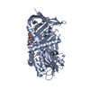
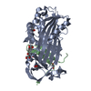
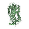
Entry Database : PDB / ID : 4bb2Title Crystal structure of cleaved corticosteroid-binding globulin in complex with progesterone (CORTICOSTEROID-BINDING ...) x 2 Keywords / / Function / homology Function Domain/homology Component
/ / / / / / / / / / / / / / / / / / / / / / / / / / / / / / / / / Biological species HOMO SAPIENS (human)Method / / / Resolution : 2.48 Å Authors Gardill, B.R. / Vogl, M.R. / Lin, H. / Hammond, G.L. / Muller, Y.A. Journal : Plos One / Year : 2012Title : Corticosteroid-Binding Globulin: Structure-Function Implications from Species DifferencesAuthors : Gardill, B.R. / Vogl, M.R. / Lin, H. / Hammond, G.L. / Muller, Y.A. History Deposition Sep 19, 2012 Deposition site / Processing site Revision 1.0 Dec 26, 2012 Provider / Type Revision 1.1 Jan 23, 2013 Group Revision 1.2 Dec 20, 2023 Group Data collection / Database references ... Data collection / Database references / Derived calculations / Other / Refinement description Category chem_comp_atom / chem_comp_bond ... chem_comp_atom / chem_comp_bond / database_2 / pdbx_database_status / pdbx_initial_refinement_model / struct_site Item _database_2.pdbx_DOI / _database_2.pdbx_database_accession ... _database_2.pdbx_DOI / _database_2.pdbx_database_accession / _pdbx_database_status.status_code_sf / _struct_site.pdbx_auth_asym_id / _struct_site.pdbx_auth_comp_id / _struct_site.pdbx_auth_seq_id Revision 1.3 Nov 20, 2024 Group / Category / pdbx_modification_feature
Show all Show less
 Movie
Movie Controller

 Yorodumi
 Open data
 Basic information
 Components
 Keywords
 Function and homology information
 HOMO SAPIENS (human)
 X-RAY DIFFRACTION /
  Authors
 Citation
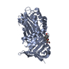
 Structure visualization
 Downloads & links
 Other downloads


 PDBj
PDBj


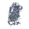
 Assembly


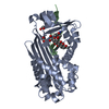
 Mass: 62.068 Da / Num. of mol.: 5 / Source method: obtained synthetically / Formula: C2H6O2
 Type: L-peptide linking / Mass: 121.158 Da / Num. of mol.: 1 / Source method: obtained synthetically / Formula: C3H7NO2S
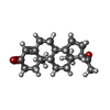
 Mass: 314.462 Da / Num. of mol.: 1 / Source method: obtained synthetically / Formula: C21H30O2
 Sample preparation
 / Beamline: 14.1 / Wavelength: 0.9184
  Processing