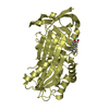
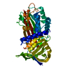
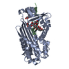
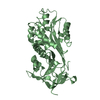
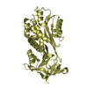
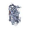
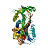
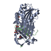
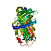
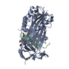
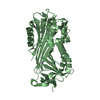
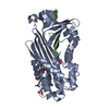
SHEET THE SHEET STRUCTURE OF THIS MOLECULE IS BIFURCATED. IN ORDER TO REPRESENT THIS FEATURE IN ... SHEET THE SHEET STRUCTURE OF THIS MOLECULE IS BIFURCATED. IN ORDER TO REPRESENT THIS FEATURE IN THE SHEET RECORDS BELOW, TWO SHEETS ARE DEFINED.
Mass: 18.015 Da / Num. of mol.: 165 / Source method: isolated from a natural source / Formula: H2O
Compound details
THE REACTIVE LOOP OF THE PROTEIN FOR BOTH CHAINS A AND B (CORRESPONDING TO UNIPROT RESIDUES 358-371) ...THE REACTIVE LOOP OF THE PROTEIN FOR BOTH CHAINS A AND B (CORRESPONDING TO UNIPROT RESIDUES 358-371) HAVE BEEN MUTATED AND ARE NOW REPLACED BY PDB RESIDUES 336-349. SEE ALSO REMARK 999.
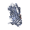
RESIDUES 336-349 OF HUMAN CBG MUTATED TO TEAAGAMFLEAIPR. THIS PROTEIN WAS CLEAVED BY THROMBIN AT ...RESIDUES 336-349 OF HUMAN CBG MUTATED TO TEAAGAMFLEAIPR. THIS PROTEIN WAS CLEAVED BY THROMBIN AT ARG349 BEFORE CRYSTALLISATION. SEE ALSO REMARK 400.
-
Experimental details
-
Experiment
Experiment
Method: X-RAY DIFFRACTION / Number of used crystals: 1
-
Sample preparation
Crystal
Density Matthews: 1.93 Å3/Da / Density % sol: 34 % / Description: NONE
 Movie
Movie Controller

 Yorodumi
 Open data
 Basic information
 Components
 Keywords
 Function and homology information
 HOMO SAPIENS (human)
 X-RAY DIFFRACTION /
  Authors
 Citation
 Structure visualization
 Downloads & links
 Other downloads


 PDBj
PDBj

 Assembly
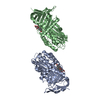


 Mass: 362.460 Da / Num. of mol.: 2 / Source method: obtained synthetically / Formula: C21H30O5
 Mass: 18.015 Da / Num. of mol.: 165 / Source method: isolated from a natural source / Formula: H2O
 Sample preparation
 Processing