 Movie
Movie Controller


+ Open data

- Basic information
| Entry | Database: PDB / ID: 4b8a | |||||||||
|---|---|---|---|---|---|---|---|---|---|---|
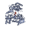
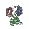
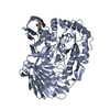
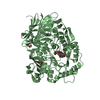
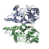
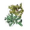
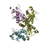
| Title | Structure of yeast NOT1 MIF4G domain co-crystallized with CAF1 | |||||||||
 Components Components |
| |||||||||
 Keywords Keywords | TRANSCRIPTION/HYDROLASE / TRANSCRIPTION-HYDROLASE COMPLEX | |||||||||
| Function / homology |  Function and homology information Function and homology informationnegative regulation of cytoplasmic mRNA processing body assembly / response to pheromone triggering conjugation with cellular fusion / pseudohyphal growth / poly(A)-specific ribonuclease / poly(A)-specific ribonuclease activity / CCR4-NOT core complex / nuclear-transcribed mRNA poly(A) tail shortening / mating projection tip / nuclear-transcribed mRNA catabolic process, deadenylation-dependent decay / ATPase activator activity ...negative regulation of cytoplasmic mRNA processing body assembly / response to pheromone triggering conjugation with cellular fusion / pseudohyphal growth / poly(A)-specific ribonuclease / poly(A)-specific ribonuclease activity / CCR4-NOT core complex / nuclear-transcribed mRNA poly(A) tail shortening / mating projection tip / nuclear-transcribed mRNA catabolic process, deadenylation-dependent decay / ATPase activator activity / transcription elongation by RNA polymerase II / P-body / positive regulation of transcription elongation by RNA polymerase II / 3'-5'-RNA exonuclease activity / molecular adaptor activity / regulation of cell cycle / negative regulation of translation / regulation of transcription by RNA polymerase II / RNA binding / metal ion binding / nucleus / cytoplasm Similarity search - Function | |||||||||
| Biological species |  | |||||||||
| Method |  X-RAY DIFFRACTION / SYNCHROTRON / MOLECULAR REPLACEMENT / Resolution: 2.4 Å X-RAY DIFFRACTION / SYNCHROTRON / MOLECULAR REPLACEMENT / Resolution: 2.4 Å | |||||||||
 Authors Authors | Basquin, J. / Conti, E. | |||||||||
 Citation Citation | Journal: Mol.Cell / Year: 2012 Title: Architecture of the Nuclease Module of the Yeast Ccr4-not Complex: The not1-Caf1-Ccr4 Interaction. Authors: Basquin, J. / Roudko, V.V. / Rode, M. / Basquin, C. / Seraphin, B. / Conti, E. | |||||||||
| History |
|
- Structure visualization
| Structure viewer | Molecule: MolmilJmol/JSmol |
|---|
- Downloads & links
-Download
| PDBx/mmCIF format | 4b8a.cif.gz | 190.3 KB | Display | PDBx/mmCIF format |
|---|---|---|---|---|
| PDB format | pdb4b8a.ent.gz | 153.2 KB | Display | PDB format |
| PDBx/mmJSON format | 4b8a.json.gz | Tree view | PDBx/mmJSON format | |
| Others |  Other downloads Other downloads |
-Validation report
| Summary document | 4b8a_validation.pdf.gz | 433.3 KB | Display | wwPDB validaton report |
|---|---|---|---|---|
| Full document | 4b8a_full_validation.pdf.gz | 438.6 KB | Display | |
| Data in XML | 4b8a_validation.xml.gz | 20.3 KB | Display | |
| Data in CIF | 4b8a_validation.cif.gz | 28.4 KB | Display | |
| Arichive directory | https://data.pdbj.org/pub/pdb/validation_reports/b8/4b8aftp://data.pdbj.org/pub/pdb/validation_reports/b8/4b8a | HTTPS FTP |
-Related structure data
| Related structure data | 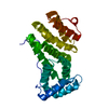 4b89C 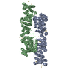 4b8bC 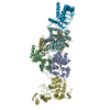 4b8cC 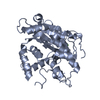 1uocS S: Starting model for refinement C: citing same article ( |
|---|---|
| Similar structure data |


-Links
 PDBj
PDBj
- Assembly
| Deposited unit | 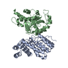
| ||||||||
|---|---|---|---|---|---|---|---|---|---|
| 1 |
| ||||||||
| Unit cell |
|
-Components
| #1: Protein | Mass: 28527.402 Da / Num. of mol.: 1 / Fragment: MIF4G, RESIDUES 755-1000 Source method: isolated from a genetically manipulated source Source: (gene. exp.)  |
|---|---|
| #2: Protein | Mass: 33153.352 Da / Num. of mol.: 1 / Fragment: NUCLEASE DOMAIN, RESIDUES 151-433 Source method: isolated from a genetically manipulated source Source: (gene. exp.) |
| #3: Water | ChemComp-HOH /  Mass: 18.015 Da / Num. of mol.: 149 / Source method: isolated from a natural source / Formula: H2O Mass: 18.015 Da / Num. of mol.: 149 / Source method: isolated from a natural source / Formula: H2O |
-Experimental details
-Experiment
| Experiment | Method: X-RAY DIFFRACTION / Number of used crystals: 1 |
|---|
- Sample preparation
| Crystal | Density Matthews: 2.53 Å3/Da / Density % sol: 51.51 % / Description: NONE |
|---|---|
| Crystal grow | pH: 8 Details: 8% (W/V) PEG 8000, 0.2 M NA MALONATE, 50 MM TRIS PH 8.0 AND 0.1% LOW MELTING AGAROSE |
-Data collection
| Diffraction | Mean temperature: 100 K |
|---|---|
| Diffraction source | Source: SYNCHROTRON / Site: SLS  / Beamline: X06DA / Wavelength: 1 / Beamline: X06DA / Wavelength: 1 |
| Detector | Type: MARRESEARCH / Detector: CCD / Date: May 19, 2010 |
| Radiation | Protocol: SINGLE WAVELENGTH / Monochromatic (M) / Laue (L): M / Scattering type: x-ray |
| Radiation wavelength | Wavelength: 1 Å / Relative weight: 1 |
| Reflection | Resolution: 2.3→47.7 Å / Num. obs: 27615 / % possible obs: 99.5 % / Observed criterion σ(I): 4.4 / Redundancy: 6 % / Rmerge(I) obs: 0.01 / Net I/σ(I): 15.3 |
| Reflection shell | Resolution: 2.3→2.45 Å / Redundancy: 5.9 % / Rmerge(I) obs: 0.04 / Mean I/σ(I) obs: 4.4 / % possible all: 96.5 |
- Processing
| Software |
| |||||||||||||||||||||||||||||||||||||||||||||||||||||||||||||||||||||||||||||
|---|---|---|---|---|---|---|---|---|---|---|---|---|---|---|---|---|---|---|---|---|---|---|---|---|---|---|---|---|---|---|---|---|---|---|---|---|---|---|---|---|---|---|---|---|---|---|---|---|---|---|---|---|---|---|---|---|---|---|---|---|---|---|---|---|---|---|---|---|---|---|---|---|---|---|---|---|---|---|
| Refinement | Method to determine structure: MOLECULAR REPLACEMENT Starting model: PDB ENTRY 1UOC Resolution: 2.4→60.14 Å / SU ML: 0.32 / σ(F): 1.99 / Phase error: 23.24 / Stereochemistry target values: ML
| |||||||||||||||||||||||||||||||||||||||||||||||||||||||||||||||||||||||||||||
| Solvent computation | Shrinkage radii: 0.9 Å / VDW probe radii: 1.11 Å / Solvent model: FLAT BULK SOLVENT MODEL / Bsol: 35.622 Å2 / ksol: 0.35 e/Å3 | |||||||||||||||||||||||||||||||||||||||||||||||||||||||||||||||||||||||||||||
| Displacement parameters |
| |||||||||||||||||||||||||||||||||||||||||||||||||||||||||||||||||||||||||||||
| Refinement step | Cycle: LAST / Resolution: 2.4→60.14 Å
| |||||||||||||||||||||||||||||||||||||||||||||||||||||||||||||||||||||||||||||
| Refine LS restraints |
| |||||||||||||||||||||||||||||||||||||||||||||||||||||||||||||||||||||||||||||
| LS refinement shell |
|
