 Movie
Movie Controller

[English] 日本語
 Yorodumi
- PDB-4auo: Crystal structure of MMP-1(E200A) in complex with a triple-helica... -
+ Open data

- Basic information
| Entry | Database: PDB / ID: 4auo | ||||||
|---|---|---|---|---|---|---|---|
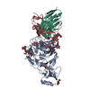
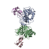
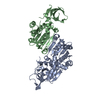
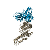
| Title | Crystal structure of MMP-1(E200A) in complex with a triple-helical collagen peptide | ||||||
 Components Components |
| ||||||
 Keywords Keywords | HYDROLASE/PEPTIDE / HYDROLASE-PEPTIDE COMPLEX | ||||||
| Function / homology |  Function and homology information Function and homology informationinterstitial collagenase / cellular response to UV-A / Basigin interactions / Activation of Matrix Metalloproteinases / Collagen degradation / collagen catabolic process / extracellular matrix disassembly / Degradation of the extracellular matrix / extracellular matrix organization / positive regulation of protein-containing complex assembly ...interstitial collagenase / cellular response to UV-A / Basigin interactions / Activation of Matrix Metalloproteinases / Collagen degradation / collagen catabolic process / extracellular matrix disassembly / Degradation of the extracellular matrix / extracellular matrix organization / positive regulation of protein-containing complex assembly / metalloendopeptidase activity / Regulation of Insulin-like Growth Factor (IGF) transport and uptake by Insulin-like Growth Factor Binding Proteins (IGFBPs) / peptidase activity / extracellular matrix / Interleukin-4 and Interleukin-13 signaling / endopeptidase activity / serine-type endopeptidase activity / proteolysis / extracellular region / zinc ion binding Similarity search - Function | ||||||
| Biological species |  HOMO SAPIENS (human) HOMO SAPIENS (human)SYNTHETIC CONSTRUCT (others) | ||||||
| Method |  X-RAY DIFFRACTION / SYNCHROTRON / MOLECULAR REPLACEMENT / Resolution: 3 Å X-RAY DIFFRACTION / SYNCHROTRON / MOLECULAR REPLACEMENT / Resolution: 3 Å | ||||||
 Authors Authors | Manka, S.W. / Carafoli, F. / Visse, R. / Bihan, D. / Raynal, N. / Farndale, R.W. / Murphy, G. / Enghild, J.J. / Hohenester, E. / Nagase, H. | ||||||
 Citation Citation | Journal: Proc.Natl.Acad.Sci.USA / Year: 2012 Title: Structural Insights Into Triple-Helical Collagen Cleavage by Matrix Metalloproteinase 1 Authors: Manka, S.W. / Carafoli, F. / Visse, R. / Bihan, D. / Raynal, N. / Farndale, R.W. / Murphy, G. / Enghild, J.J. / Hohenester, E. / Nagase, H. | ||||||
| History |
|
- Structure visualization
| Structure viewer | Molecule: MolmilJmol/JSmol |
|---|
- Downloads & links
-Download
| PDBx/mmCIF format | 4auo.cif.gz | 197.3 KB | Display | PDBx/mmCIF format |
|---|---|---|---|---|
| PDB format | pdb4auo.ent.gz | 157.9 KB | Display | PDB format |
| PDBx/mmJSON format | 4auo.json.gz | Tree view | PDBx/mmJSON format | |
| Others |  Other downloads Other downloads |
-Validation report
| Arichive directory | https://data.pdbj.org/pub/pdb/validation_reports/au/4auoftp://data.pdbj.org/pub/pdb/validation_reports/au/4auo | HTTPS FTP |
|---|
-Related structure data
| Related structure data |  2cltS S: Starting model for refinement |
|---|---|
| Similar structure data |


-Links
 PDBj
PDBj


- Assembly
| Deposited unit | 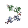
| ||||||||
|---|---|---|---|---|---|---|---|---|---|
| 1 | 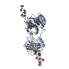
| ||||||||
| 2 | 
| ||||||||
| Unit cell |
|
-Components
| #1: Protein | Mass: 42230.070 Da / Num. of mol.: 2 / Mutation: YES Source method: isolated from a genetically manipulated source Source: (gene. exp.) HOMO SAPIENS (human) / Production host:  #2: Protein/peptide | Mass: 3783.110 Da / Num. of mol.: 6 / Source method: obtained synthetically / Source: (synth.) SYNTHETIC CONSTRUCT (others) #3: Chemical | ChemComp-CA /   Mass: 40.078 Da / Num. of mol.: 8 / Source method: obtained synthetically / Formula: Ca Mass: 40.078 Da / Num. of mol.: 8 / Source method: obtained synthetically / Formula: Ca#4: Chemical | ChemComp-ZN /   Mass: 65.409 Da / Num. of mol.: 4 / Source method: obtained synthetically / Formula: Zn Mass: 65.409 Da / Num. of mol.: 4 / Source method: obtained synthetically / Formula: Zn#5: Water | ChemComp-HOH / |  Mass: 18.015 Da / Num. of mol.: 7 / Source method: isolated from a natural source / Formula: H2O Mass: 18.015 Da / Num. of mol.: 7 / Source method: isolated from a natural source / Formula: H2OCompound details | ENGINEERED | Sequence details | THE CRYSTALLIS | |
|---|
-Experimental details
-Experiment
| Experiment | Method: X-RAY DIFFRACTION / Number of used crystals: 1 |
|---|
- Sample preparation
| Crystal | Density Matthews: 2.87 Å3/Da / Density % sol: 57.12 % / Description: NONE |
|---|---|
| Crystal grow | pH: 8 / Details: pH 8 |
-Data collection
| Diffraction | Mean temperature: 100 K |
|---|---|
| Diffraction source | Source: SYNCHROTRON / Site: Diamond  / Beamline: I24 / Wavelength: 0.9778 / Beamline: I24 / Wavelength: 0.9778 |
| Detector | Type: DECTRIS PILATUS 6M / Detector: PIXEL / Date: Dec 13, 2010 |
| Radiation | Protocol: SINGLE WAVELENGTH / Monochromatic (M) / Laue (L): M / Scattering type: x-ray |
| Radiation wavelength | Wavelength: 0.9778 Å / Relative weight: 1 |
| Reflection | Resolution: 3→20 Å / Num. obs: 20982 / % possible obs: 86.8 % / Observed criterion σ(I): 0 / Redundancy: 1.7 % / Rmerge(I) obs: 0.13 / Net I/σ(I): 5 |
| Reflection shell | Resolution: 3→3.16 Å / Redundancy: 1.6 % / Rmerge(I) obs: 0.4 / Mean I/σ(I) obs: 2.2 / % possible all: 77.9 |
- Processing
| Software |
| ||||||||||||||||||||||||||||||||||||||||||||||||||||||||||||
|---|---|---|---|---|---|---|---|---|---|---|---|---|---|---|---|---|---|---|---|---|---|---|---|---|---|---|---|---|---|---|---|---|---|---|---|---|---|---|---|---|---|---|---|---|---|---|---|---|---|---|---|---|---|---|---|---|---|---|---|---|---|
| Refinement | Method to determine structure: MOLECULAR REPLACEMENT Starting model: PDB ENTRY 2CLT Resolution: 3→20 Å / Data cutoff high absF: 10000 / Data cutoff low absF: 0 / Cross valid method: THROUGHOUT / σ(F): 0 / Stereochemistry target values: MAXIMUM LIKELIHOOD Details: THERE ARE TWO COMPLEXES IN THE ASYMMETRIC UNIT (CHAINS ACDE AND BFGH). THEY DIFFER ONLY IN THE ENDS OF THE COLLAGEN MOLECULES.
| ||||||||||||||||||||||||||||||||||||||||||||||||||||||||||||
| Solvent computation | Solvent model: FLAT MODEL / Bsol: 13.979 Å2 / ksol: 0.35 e/Å3 | ||||||||||||||||||||||||||||||||||||||||||||||||||||||||||||
| Displacement parameters |
| ||||||||||||||||||||||||||||||||||||||||||||||||||||||||||||
| Refinement step | Cycle: LAST / Resolution: 3→20 Å
| ||||||||||||||||||||||||||||||||||||||||||||||||||||||||||||
| Refine LS restraints |
| ||||||||||||||||||||||||||||||||||||||||||||||||||||||||||||
| Xplor file |
|
