 Movie
Movie Controller

[English] 日本語
 Yorodumi
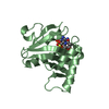
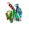
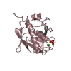
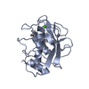
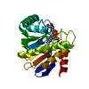
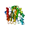
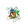
- PDB-1cgf: CRYSTAL STRUCTURES OF RECOMBINANT 19-KDA HUMAN FIBROBLAST COLLAGE... -
+ Open data

- Basic information
| Entry | Database: PDB / ID: 1cgf | ||||||
|---|---|---|---|---|---|---|---|
| Title | CRYSTAL STRUCTURES OF RECOMBINANT 19-KDA HUMAN FIBROBLAST COLLAGENASE COMPLEXED TO ITSELF | ||||||
 Components Components | FIBROBLAST COLLAGENASE | ||||||
 Keywords Keywords | HYDROLASE (METALLOPROTEASE) | ||||||
| Function / homology |  Function and homology information Function and homology informationinterstitial collagenase / cellular response to UV-A / Basigin interactions / Activation of Matrix Metalloproteinases / Collagen degradation / collagen catabolic process / extracellular matrix disassembly / Degradation of the extracellular matrix / extracellular matrix organization / positive regulation of protein-containing complex assembly ...interstitial collagenase / cellular response to UV-A / Basigin interactions / Activation of Matrix Metalloproteinases / Collagen degradation / collagen catabolic process / extracellular matrix disassembly / Degradation of the extracellular matrix / extracellular matrix organization / positive regulation of protein-containing complex assembly / metalloendopeptidase activity / Regulation of Insulin-like Growth Factor (IGF) transport and uptake by Insulin-like Growth Factor Binding Proteins (IGFBPs) / peptidase activity / extracellular matrix / Interleukin-4 and Interleukin-13 signaling / endopeptidase activity / serine-type endopeptidase activity / proteolysis / extracellular region / zinc ion binding Similarity search - Function | ||||||
| Biological species |  Homo sapiens (human) Homo sapiens (human) | ||||||
| Method |  X-RAY DIFFRACTION / Resolution: 2.1 Å X-RAY DIFFRACTION / Resolution: 2.1 Å | ||||||
 Authors Authors | Lovejoy, B. / Hassell, A.M. / Luther, M.A. / Weigl, D. / Jordan, S.R. | ||||||
 Citation Citation | Journal: Biochemistry / Year: 1994 Title: Crystal structures of recombinant 19-kDa human fibroblast collagenase complexed to itself. Authors: Lovejoy, B. / Hassell, A.M. / Luther, M.A. / Weigl, D. / Jordan, S.R. #1: Journal: Science / Year: 1994Title: Structure of the Catalytic Domain of Fibroblast Collagenase Complexed with an Inhibitor Authors: Lovejoy, B. / Cleasby, A. / Hassell, A.M. / Longley, K. / Luther, M.A. / Weigl, D. / Mcgeehan, G. / Mcelroy, A.B. / Drewry, D. / Lambert, M.H. / Jordan, S.R. | ||||||
| History |
|
- Structure visualization
| Structure viewer | Molecule: MolmilJmol/JSmol |
|---|
- Downloads & links
-Download
| PDBx/mmCIF format | 1cgf.cif.gz | 99.2 KB | Display | PDBx/mmCIF format |
|---|---|---|---|---|
| PDB format | pdb1cgf.ent.gz | 76.5 KB | Display | PDB format |
| PDBx/mmJSON format | 1cgf.json.gz | Tree view | PDBx/mmJSON format | |
| Others |  Other downloads Other downloads |
-Validation report
| Arichive directory | https://data.pdbj.org/pub/pdb/validation_reports/cg/1cgfftp://data.pdbj.org/pub/pdb/validation_reports/cg/1cgf | HTTPS FTP |
|---|
-Related structure data


-Links
 PDBj
PDBj


- Assembly
| Deposited unit | 
| ||||||||
|---|---|---|---|---|---|---|---|---|---|
| 1 | 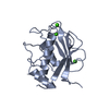
| ||||||||
| 2 | 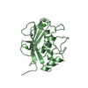
| ||||||||
| Unit cell |
|
-Components
| #1: Protein | Mass: 18158.775 Da / Num. of mol.: 2 Source method: isolated from a genetically manipulated source Source: (gene. exp.) Homo sapiens (human) / Gene: CDNA / References: UniProt: P03956, interstitial collagenase#2: Chemical | ChemComp-ZN /   Mass: 65.409 Da / Num. of mol.: 4 / Source method: obtained synthetically / Formula: Zn Mass: 65.409 Da / Num. of mol.: 4 / Source method: obtained synthetically / Formula: Zn#3: Chemical | ChemComp-CA /   Mass: 40.078 Da / Num. of mol.: 6 / Source method: obtained synthetically / Formula: Ca Mass: 40.078 Da / Num. of mol.: 6 / Source method: obtained synthetically / Formula: Ca#4: Water | ChemComp-HOH / |  Mass: 18.015 Da / Num. of mol.: 181 / Source method: isolated from a natural source / Formula: H2O Mass: 18.015 Da / Num. of mol.: 181 / Source method: isolated from a natural source / Formula: H2OSequence details | THE SEQUENCE WAS DERIVED FROM THE CDNA SEQUENCE OF G. I. GOLDBERG, ET AL., J. BIOL. CHEM. 261: 6600- ...THE SEQUENCE WAS DERIVED FROM THE CDNA SEQUENCE OF G. I. GOLDBERG, ET AL., J. BIOL. CHEM. 261: 6600-660 (1986), AND DESCRIBED IN THE JRNL REFERENCE ABOVE. N-TERMINAL MASS SPECTROMET | |
|---|
-Experimental details
-Experiment
| Experiment | Method: X-RAY DIFFRACTION |
|---|
- Sample preparation
| Crystal | Density Matthews: 2.33 Å3/Da / Density % sol: 47.26 % | |||||||||||||||||||||||||
|---|---|---|---|---|---|---|---|---|---|---|---|---|---|---|---|---|---|---|---|---|---|---|---|---|---|---|
| Crystal grow | *PLUS Temperature: 4 ℃ / Method: vapor diffusion, hanging drop / PH range low: 9 / PH range high: 7 | |||||||||||||||||||||||||
| Components of the solutions | *PLUS
|
-Data collection
| Radiation | Scattering type: x-ray |
|---|---|
| Radiation wavelength | Relative weight: 1 |
| Reflection | *PLUS Highest resolution: 2.1 Å / Num. obs: 17088 / % possible obs: 86.7 % / Rmerge(I) obs: 0.062 |
- Processing
| Software |
| ||||||||||||||||||||||||||||||||||||||||||||||||||||||||||||
|---|---|---|---|---|---|---|---|---|---|---|---|---|---|---|---|---|---|---|---|---|---|---|---|---|---|---|---|---|---|---|---|---|---|---|---|---|---|---|---|---|---|---|---|---|---|---|---|---|---|---|---|---|---|---|---|---|---|---|---|---|---|
| Refinement | Resolution: 2.1→7 Å / σ(F): 1 /
| ||||||||||||||||||||||||||||||||||||||||||||||||||||||||||||
| Refinement step | Cycle: LAST / Resolution: 2.1→7 Å
| ||||||||||||||||||||||||||||||||||||||||||||||||||||||||||||
| Refine LS restraints |
| ||||||||||||||||||||||||||||||||||||||||||||||||||||||||||||
| Refinement | *PLUS | ||||||||||||||||||||||||||||||||||||||||||||||||||||||||||||
| Solvent computation | *PLUS | ||||||||||||||||||||||||||||||||||||||||||||||||||||||||||||
| Displacement parameters | *PLUS | ||||||||||||||||||||||||||||||||||||||||||||||||||||||||||||
| Refine LS restraints | *PLUS Type: x_angle_d / Dev ideal: 2.06 |
