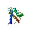
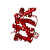
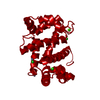
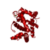
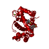
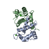
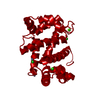
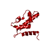
Entry Database : PDB / ID : 4aqjTitle Structure of human S100A7 D24G bound to zinc and calcium PROTEIN S100-A7 Keywords / / / Function / homology Function Domain/homology Component
/ / / / / / / / / / / / / / / / / / / / / / / / / / / / / / / / / / / / / / / / / / / / / / / / / / / / / / / / / / Biological species HOMO SAPIENS (human)Method / / / Resolution : 1.6 Å Authors Murray, J.I. / Tonkin, M.L. / Whiting, A.L. / Peng, F. / Farnell, B. / Hof, F. / Boulanger, M.J. Journal : Bmc Struct.Biol. / Year : 2012Title : Structural Characterization of S100A15 Reveals a Novel Zinc Coordination Site Among S100 Proteins and Altered Surface Chemistry with Functional Implications for Receptor Binding.Authors : Murray, J.I. / Tonkin, M.L. / Whiting, A.L. / Peng, F. / Farnell, B. / Cullen, J.T. / Hof, F. / Boulanger, M.J. History Deposition Apr 17, 2012 Deposition site / Processing site Revision 1.0 Oct 17, 2012 Provider / Type Revision 1.1 Nov 20, 2024 Group Data collection / Database references ... Data collection / Database references / Derived calculations / Other / Structure summary Category chem_comp_atom / chem_comp_bond ... chem_comp_atom / chem_comp_bond / database_2 / pdbx_database_status / pdbx_entry_details / pdbx_modification_feature / pdbx_struct_conn_angle / struct_conn / struct_site Item _database_2.pdbx_DOI / _database_2.pdbx_database_accession ... _database_2.pdbx_DOI / _database_2.pdbx_database_accession / _pdbx_database_status.status_code_sf / _pdbx_entry_details.has_protein_modification / _pdbx_struct_conn_angle.ptnr1_auth_comp_id / _pdbx_struct_conn_angle.ptnr1_auth_seq_id / _pdbx_struct_conn_angle.ptnr1_label_asym_id / _pdbx_struct_conn_angle.ptnr1_label_atom_id / _pdbx_struct_conn_angle.ptnr1_label_comp_id / _pdbx_struct_conn_angle.ptnr1_label_seq_id / _pdbx_struct_conn_angle.ptnr1_symmetry / _pdbx_struct_conn_angle.ptnr2_auth_comp_id / _pdbx_struct_conn_angle.ptnr2_auth_seq_id / _pdbx_struct_conn_angle.ptnr2_label_asym_id / _pdbx_struct_conn_angle.ptnr2_label_atom_id / _pdbx_struct_conn_angle.ptnr2_label_comp_id / _pdbx_struct_conn_angle.ptnr3_auth_comp_id / _pdbx_struct_conn_angle.ptnr3_auth_seq_id / _pdbx_struct_conn_angle.ptnr3_label_asym_id / _pdbx_struct_conn_angle.ptnr3_label_atom_id / _pdbx_struct_conn_angle.ptnr3_label_comp_id / _pdbx_struct_conn_angle.ptnr3_label_seq_id / _pdbx_struct_conn_angle.ptnr3_symmetry / _pdbx_struct_conn_angle.value / _struct_conn.pdbx_dist_value / _struct_conn.ptnr1_auth_comp_id / _struct_conn.ptnr1_auth_seq_id / _struct_conn.ptnr1_label_asym_id / _struct_conn.ptnr1_label_atom_id / _struct_conn.ptnr1_label_comp_id / _struct_conn.ptnr1_label_seq_id / _struct_conn.ptnr1_symmetry / _struct_conn.ptnr2_auth_comp_id / _struct_conn.ptnr2_auth_seq_id / _struct_conn.ptnr2_label_asym_id / _struct_conn.ptnr2_label_atom_id / _struct_conn.ptnr2_label_comp_id / _struct_conn.ptnr2_label_seq_id / _struct_conn.ptnr2_symmetry / _struct_site.pdbx_auth_asym_id / _struct_site.pdbx_auth_comp_id / _struct_site.pdbx_auth_seq_id
Show all Show less
 Open data
 Basic information
 Components
 Keywords
 Function and homology information
 HOMO SAPIENS (human)
 X-RAY DIFFRACTION /
  SYNCHROTRON /
  MOLECULAR REPLACEMENT / Resolution: 1.6 Å
  Authors
 Citation
 Journal: Bmc Struct.Biol. / Year: 2012
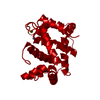
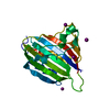
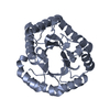
 Structure visualization
 Molmil
 Jmol/JSmol
 Downloads & links
 Download
 4aqj.cif.gz
 PDBx/mmCIF format
 pdb4aqj.ent.gz
 PDB format
 4aqj.json.gz
 PDBx/mmJSON format
 Other downloads
 https://data.pdbj.org/pub/pdb/validation_reports/aq/4aqj
 ftp://data.pdbj.org/pub/pdb/validation_reports/aq/4aqj
 Links
Links Assembly
 Components
 HOMO SAPIENS (human) / Production host:
 X-RAY DIFFRACTION
 Sample preparation
 SYNCHROTRON / Site:
  SSRL
SSRL  / Beamline: BL9-2 / Wavelength: 0.9794
  Processing
 MOLECULAR REPLACEMENT
 Movie
Movie Controller









 PDBj
PDBj


