 Movie
Movie Controller

[English] 日本語
 Yorodumi
- PDB-2zvr: Crystal structure of a D-tagatose 3-epimerase-related protein fro... -
+ Open data

- Basic information
| Entry | Database: PDB / ID: 2zvr | ||||||
|---|---|---|---|---|---|---|---|
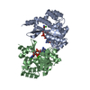
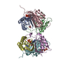
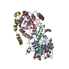
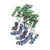
| Title | Crystal structure of a D-tagatose 3-epimerase-related protein from Thermotoga maritima | ||||||
 Components Components | Uncharacterized protein TM_0416 | ||||||
 Keywords Keywords | ISOMERASE / hyperthermophile / Thermotoga maritima / ketohexose 3-epimerase / D-tagatose 3-epimerase | ||||||
| Function / homology |  Function and homology information Function and homology informationIsomerases; Intramolecular oxidoreductases; Interconverting aldoses and ketoses, and related compounds / Isomerases; Racemases and epimerases; Acting on carbohydrates and derivatives / inositol metabolic process / isomerase activity / metal ion binding Similarity search - Function | ||||||
| Biological species |   Thermotoga maritima (bacteria) Thermotoga maritima (bacteria) | ||||||
| Method |  X-RAY DIFFRACTION / SIRAS / Resolution: 2.2 Å X-RAY DIFFRACTION / SIRAS / Resolution: 2.2 Å | ||||||
 Authors Authors | Sakuraba, H. / Ohshima, T. | ||||||
 Citation Citation | Journal: Acta Crystallogr.,Sect.F / Year: 2009 Title: Structure of a D-tagatose 3-epimerase-related protein from the hyperthermophilic bacterium Thermotoga maritima Authors: Sakuraba, H. / Yoneda, K. / Satomura, T. / Kawakami, R. / Ohshima, T. | ||||||
| History |
|
- Structure visualization
| Structure viewer | Molecule: MolmilJmol/JSmol |
|---|
- Downloads & links
-Download
| PDBx/mmCIF format | 2zvr.cif.gz | 117.5 KB | Display | PDBx/mmCIF format |
|---|---|---|---|---|
| PDB format | pdb2zvr.ent.gz | 90.6 KB | Display | PDB format |
| PDBx/mmJSON format | 2zvr.json.gz | Tree view | PDBx/mmJSON format | |
| Others |  Other downloads Other downloads |
-Validation report
| Arichive directory | https://data.pdbj.org/pub/pdb/validation_reports/zv/2zvrftp://data.pdbj.org/pub/pdb/validation_reports/zv/2zvr | HTTPS FTP |
|---|
-Related structure data
| Similar structure data |
|---|


-Links
 PDBj
PDBj
- Assembly
| Deposited unit | 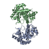
| ||||||||
|---|---|---|---|---|---|---|---|---|---|
| 1 |
| ||||||||
| 2 | 
| ||||||||
| 3 | 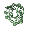
| ||||||||
| Unit cell |
|
-Components
| #1: Protein | Mass: 32677.482 Da / Num. of mol.: 2 Source method: isolated from a genetically manipulated source Source: (gene. exp.) Thermotoga maritima (bacteria) / Gene: TM_0416 / Plasmid: pET-15b / Production host: #2: Water | ChemComp-HOH / |  Mass: 18.015 Da / Num. of mol.: 231 / Source method: isolated from a natural source / Formula: H2O Mass: 18.015 Da / Num. of mol.: 231 / Source method: isolated from a natural source / Formula: H2O |
|---|
-Experimental details
-Experiment
| Experiment | Method: X-RAY DIFFRACTION / Number of used crystals: 1 |
|---|
- Sample preparation
| Crystal | Density Matthews: 2.31 Å3/Da / Density % sol: 46.8 % |
|---|---|
| Crystal grow | Temperature: 293 K / Method: vapor diffusion, sitting drop / pH: 6 Details: 20-30% PEG 200, 5% PEG 1000, 0.1M MES, pH 6, VAPOR DIFFUSION, SITTING DROP, temperature 293K |
-Data collection
| Diffraction | Mean temperature: 100 K |
|---|---|
| Diffraction source | Source: ROTATING ANODE / Type: RIGAKU MICROMAX-007 / Wavelength: 1.5418 Å |
| Detector | Type: RIGAKU RAXIS VII / Detector: IMAGE PLATE / Date: Sep 3, 2008 |
| Radiation | Monochromator: SI / Protocol: SINGLE WAVELENGTH / Monochromatic (M) / Laue (L): M / Scattering type: x-ray |
| Radiation wavelength | Wavelength: 1.5418 Å / Relative weight: 1 |
| Reflection | Resolution: 2.17→37.63 Å / Num. obs: 29673 / Observed criterion σ(F): 0 / Observed criterion σ(I): 0 / Biso Wilson estimate: 24.1 Å2 |
| Reflection shell | Resolution: 2.17→2.25 Å |
- Processing
| Software |
| |||||||||||||||||||||||||
|---|---|---|---|---|---|---|---|---|---|---|---|---|---|---|---|---|---|---|---|---|---|---|---|---|---|---|
| Refinement | Method to determine structure: SIRAS / Resolution: 2.2→37.6 Å / Rfactor Rfree error: 0.004 / Data cutoff high absF: 1207194.3 / Data cutoff low absF: 0 / Isotropic thermal model: RESTRAINED / Cross valid method: THROUGHOUT / σ(F): 0 / σ(I): 0 / Stereochemistry target values: Engh & Huber / Details: BULK SOLVENT MODEL USED
| |||||||||||||||||||||||||
| Solvent computation | Solvent model: FLAT MODEL / Bsol: 46.0099 Å2 / ksol: 0.4 e/Å3 | |||||||||||||||||||||||||
| Displacement parameters | Biso mean: 20.6 Å2
| |||||||||||||||||||||||||
| Refine analyze |
| |||||||||||||||||||||||||
| Refinement step | Cycle: LAST / Resolution: 2.2→37.6 Å
| |||||||||||||||||||||||||
| Refine LS restraints |
| |||||||||||||||||||||||||
| LS refinement shell | Resolution: 2.17→2.31 Å / Rfactor Rfree error: 0.011 / Total num. of bins used: 6
| |||||||||||||||||||||||||
| Xplor file |
|
