 Movie
Movie Controller

[English] 日本語
 Yorodumi
- PDB-6aq5: X-ray crystal structure of Erythrina crista-galli lectin in compl... -
+ Open data

- Basic information
| Entry | Database: PDB / ID: 6aq5 | |||||||||
|---|---|---|---|---|---|---|---|---|---|---|
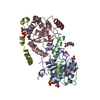
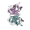
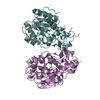
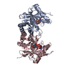
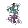
| Title | X-ray crystal structure of Erythrina crista-galli lectin in complex with epilactose | |||||||||
 Components Components | Lectin | |||||||||
 Keywords Keywords | SUGAR BINDING PROTEIN / carbohydrate binding protein / lectin / epilactose | |||||||||
| Function / homology |  Function and homology information Function and homology information | |||||||||
| Biological species |  Erythrina crista-galli (cockspur coraltree) Erythrina crista-galli (cockspur coraltree) | |||||||||
| Method |  X-RAY DIFFRACTION / MOLECULAR REPLACEMENT / Resolution: 2.194 Å X-RAY DIFFRACTION / MOLECULAR REPLACEMENT / Resolution: 2.194 Å | |||||||||
 Authors Authors | Gerlits, O. / Woods, R.J. | |||||||||
 Citation Citation | Journal: To Be Published Title: X-ray crystal structure of Erythrina crista-galli lectin in complex with epilactose Authors: Gerlits, O. / Woods, R.J. / Sood, A. | |||||||||
| History |
|
- Structure visualization
| Structure viewer | Molecule: MolmilJmol/JSmol |
|---|
- Downloads & links
-Download
| PDBx/mmCIF format | 6aq5.cif.gz | 118.5 KB | Display | PDBx/mmCIF format |
|---|---|---|---|---|
| PDB format | pdb6aq5.ent.gz | 89.4 KB | Display | PDB format |
| PDBx/mmJSON format | 6aq5.json.gz | Tree view | PDBx/mmJSON format | |
| Others |  Other downloads Other downloads |
-Validation report
| Arichive directory | https://data.pdbj.org/pub/pdb/validation_reports/aq/6aq5ftp://data.pdbj.org/pub/pdb/validation_reports/aq/6aq5 | HTTPS FTP |
|---|
-Related structure data
| Related structure data | 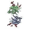 1uzyS S: Starting model for refinement |
|---|---|
| Similar structure data |


-Links
 PDBj
PDBj

- Assembly
| Deposited unit | 
| ||||||||
|---|---|---|---|---|---|---|---|---|---|
| 1 |
| ||||||||
| Unit cell |
|
-Components
-Protein , 1 types, 2 molecules AB
| #1: Protein | Mass: 26236.234 Da / Num. of mol.: 2 / Source method: isolated from a natural source Source: (natural) Erythrina crista-galli (cockspur coraltree)References: UniProt: Q6YD91 |
|---|
-Sugars , 3 types, 4 molecules 
| #2: Polysaccharide | Source method: isolated from a genetically manipulated source #3: Polysaccharide | beta-D-xylopyranose-(1-2)-[alpha-D-mannopyranose-(1-3)]beta-D-mannopyranose-(1-4)-2-acetamido-2- ...beta-D-xylopyranose-(1-2)-[alpha-D-mannopyranose-(1-3)]beta-D-mannopyranose-(1-4)-2-acetamido-2-deoxy-beta-D-glucopyranose-(1-4)-[alpha-L-fucopyranose-(1-3)]2-acetamido-2-deoxy-beta-D-glucopyranose | Source method: isolated from a genetically manipulated source #7: Sugar | ChemComp-NAG / |  Type: D-saccharide, beta linking / Mass: 221.208 Da / Num. of mol.: 1 Type: D-saccharide, beta linking / Mass: 221.208 Da / Num. of mol.: 1Source method: isolated from a genetically manipulated source Formula: C8H15NO6 |
|---|
-Non-polymers , 4 types, 297 molecules 


| #4: Chemical |  Mass: 54.938 Da / Num. of mol.: 2 Mass: 54.938 Da / Num. of mol.: 2Source method: isolated from a genetically manipulated source Formula: Mn #5: Chemical |  Mass: 40.078 Da / Num. of mol.: 2 Mass: 40.078 Da / Num. of mol.: 2Source method: isolated from a genetically manipulated source Formula: Ca #6: Chemical | ChemComp-NA / |  Mass: 22.990 Da / Num. of mol.: 1 Mass: 22.990 Da / Num. of mol.: 1Source method: isolated from a genetically manipulated source Formula: Na #8: Water | ChemComp-HOH / | Mass: 18.015 Da / Num. of mol.: 292 / Source method: isolated from a natural source / Formula: H2O |
|---|
-Details
| Has protein modification | Y |
|---|
-Experimental details
-Experiment
| Experiment | Method: X-RAY DIFFRACTION / Number of used crystals: 1 |
|---|
- Sample preparation
| Crystal | Density Matthews: 4.1 Å3/Da / Density % sol: 69.98 % |
|---|---|
| Crystal grow | Temperature: 293.15 K / Method: vapor diffusion, sitting drop / pH: 7.5 / Details: PEG3350, calcium acetate, HEPES, pH 7.5 |
-Data collection
| Diffraction | Mean temperature: 100 K |
|---|---|
| Diffraction source | Source: ROTATING ANODE / Type: RIGAKU MICROMAX-007 HF / Wavelength: 1.54 Å |
| Detector | Type: RIGAKU RAXIS IV++ / Detector: IMAGE PLATE / Date: Sep 29, 2016 |
| Radiation | Protocol: SINGLE WAVELENGTH / Monochromatic (M) / Laue (L): M / Scattering type: x-ray |
| Radiation wavelength | Wavelength: 1.54 Å / Relative weight: 1 |
| Reflection | Resolution: 2.2→40 Å / Num. obs: 42764 / % possible obs: 98.8 % / Redundancy: 3.3 % / Rsym value: 0.085 / Net I/σ(I): 13.1 |
| Reflection shell | Resolution: 2.2→2.28 Å / Rsym value: 0.496 |
- Processing
| Software |
| ||||||||||||||||||||||||||||||||||||||||||||||||||||||||||||||||||||||||||||||||||||||||||||||||||
|---|---|---|---|---|---|---|---|---|---|---|---|---|---|---|---|---|---|---|---|---|---|---|---|---|---|---|---|---|---|---|---|---|---|---|---|---|---|---|---|---|---|---|---|---|---|---|---|---|---|---|---|---|---|---|---|---|---|---|---|---|---|---|---|---|---|---|---|---|---|---|---|---|---|---|---|---|---|---|---|---|---|---|---|---|---|---|---|---|---|---|---|---|---|---|---|---|---|---|---|
| Refinement | Method to determine structure: MOLECULAR REPLACEMENT Starting model: PDB entry 1UZY Resolution: 2.194→35.171 Å / SU ML: 0.27 / Cross valid method: FREE R-VALUE / σ(F): 1.36 / Phase error: 21.47 / Stereochemistry target values: ML
| ||||||||||||||||||||||||||||||||||||||||||||||||||||||||||||||||||||||||||||||||||||||||||||||||||
| Solvent computation | Shrinkage radii: 0.9 Å / VDW probe radii: 1.11 Å / Solvent model: FLAT BULK SOLVENT MODEL | ||||||||||||||||||||||||||||||||||||||||||||||||||||||||||||||||||||||||||||||||||||||||||||||||||
| Refinement step | Cycle: LAST / Resolution: 2.194→35.171 Å
| ||||||||||||||||||||||||||||||||||||||||||||||||||||||||||||||||||||||||||||||||||||||||||||||||||
| Refine LS restraints |
| ||||||||||||||||||||||||||||||||||||||||||||||||||||||||||||||||||||||||||||||||||||||||||||||||||
| LS refinement shell |
|
