 Movie
Movie Controller

[English] 日本語
 Yorodumi
- PDB-1fay: WINGED BEAN ACIDIC LECTIN COMPLEXED WITH METHYL-ALPHA-D-GALACTOSE... -
+ Open data

- Basic information
| Entry | Database: PDB / ID: 1fay | |||||||||
|---|---|---|---|---|---|---|---|---|---|---|
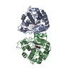
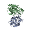
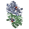
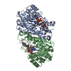
| Title | WINGED BEAN ACIDIC LECTIN COMPLEXED WITH METHYL-ALPHA-D-GALACTOSE (MONOCLINIC FORM) | |||||||||
 Components Components | ACIDIC LECTIN | |||||||||
 Keywords Keywords | SUGAR BINDING PROTEIN / LEGUME LECTIN / GLYCOSYLATED PROTEIN / H-ANTIGENIC SPECIFICITY / AGGLUTININ | |||||||||
| Function / homology |  Function and homology information Function and homology information | |||||||||
| Biological species |   Psophocarpus tetragonolobus (winged bean) Psophocarpus tetragonolobus (winged bean) | |||||||||
| Method |  X-RAY DIFFRACTION / MOLECULAR REPLACEMENT / Resolution: 3.3 Å X-RAY DIFFRACTION / MOLECULAR REPLACEMENT / Resolution: 3.3 Å | |||||||||
 Authors Authors | Manoj, N. / Srinivas, V.R. / Surolia, A. / Vijayan, M. / Suguna, K. | |||||||||
 Citation Citation | Journal: J.Mol.Biol. / Year: 2000 Title: Carbohydrate specificity and salt-bridge mediated conformational change in acidic winged bean agglutinin. Authors: Manoj, N. / Srinivas, V.R. / Surolia, A. / Vijayan, M. / Suguna, K. #1: Journal: Acta Crystallogr.,Sect.D / Year: 1999Title: Crystallization and Preliminary Crystallographic Analysis of Winged Bean Acidic Lectin Authors: Manoj, N. / Srinivas, V.R. / Satish, B. / Singha, N.C. / Suguna, K. | |||||||||
| History |
|
- Structure visualization
| Structure viewer | Molecule: MolmilJmol/JSmol |
|---|
- Downloads & links
-Download
| PDBx/mmCIF format | 1fay.cif.gz | 337.7 KB | Display | PDBx/mmCIF format |
|---|---|---|---|---|
| PDB format | pdb1fay.ent.gz | 275.2 KB | Display | PDB format |
| PDBx/mmJSON format | 1fay.json.gz | Tree view | PDBx/mmJSON format | |
| Others |  Other downloads Other downloads |
-Validation report
| Arichive directory | https://data.pdbj.org/pub/pdb/validation_reports/fa/1fayftp://data.pdbj.org/pub/pdb/validation_reports/fa/1fay | HTTPS FTP |
|---|
-Related structure data
| Related structure data |  1f9kSC S: Starting model for refinement C: citing same article ( |
|---|---|
| Similar structure data |


-Links
 PDBj
PDBj

- Assembly
| Deposited unit | 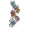
| ||||||||
|---|---|---|---|---|---|---|---|---|---|
| 1 | 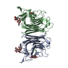
| ||||||||
| 2 | 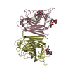
| ||||||||
| 3 | 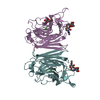
| ||||||||
| 4 | 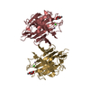
| ||||||||
| Unit cell |
| ||||||||
| Details | the biological assembly is a dimer |
-Components
-Protein , 1 types, 8 molecules ABCDEFGH
| #1: Protein | Mass: 26390.123 Da / Num. of mol.: 8 / Source method: isolated from a natural source / Details: WINGED BEAN, PSOPHOCARPUS TETRAGONOLOBUS, SEEDS / Source: (natural) Psophocarpus tetragonolobus (winged bean) / Organ: LEGUMINOUS SEEDS / References: GenBank: 6018681, UniProt: Q9SM56*PLUS |
|---|
-Sugars , 2 types, 14 molecules 
| #2: Polysaccharide | 2-acetamido-2-deoxy-beta-D-glucopyranose-(1-4)-2-acetamido-2-deoxy-beta-D-glucopyranose Source method: isolated from a genetically manipulated source #3: Sugar | ChemComp-AMG /  Type: D-saccharide / Mass: 194.182 Da / Num. of mol.: 8 / Source method: obtained synthetically / Formula: C7H14O6 Type: D-saccharide / Mass: 194.182 Da / Num. of mol.: 8 / Source method: obtained synthetically / Formula: C7H14O6 |
|---|
-Non-polymers , 3 types, 96 molecules 


| #4: Chemical | ChemComp-MN /  Mass: 54.938 Da / Num. of mol.: 8 / Source method: obtained synthetically / Formula: Mn Mass: 54.938 Da / Num. of mol.: 8 / Source method: obtained synthetically / Formula: Mn#5: Chemical | ChemComp-CA /  Mass: 40.078 Da / Num. of mol.: 8 / Source method: obtained synthetically / Formula: Ca Mass: 40.078 Da / Num. of mol.: 8 / Source method: obtained synthetically / Formula: Ca#6: Water | ChemComp-HOH / | Mass: 18.015 Da / Num. of mol.: 80 / Source method: isolated from a natural source / Formula: H2O |
|---|
-Details
| Has protein modification | Y |
|---|
-Experimental details
-Experiment
| Experiment | Method: X-RAY DIFFRACTION / Number of used crystals: 1 |
|---|
- Sample preparation
| Crystal | Density Matthews: 2.77 Å3/Da / Density % sol: 56 % | ||||||||||||||||||||||||||||||
|---|---|---|---|---|---|---|---|---|---|---|---|---|---|---|---|---|---|---|---|---|---|---|---|---|---|---|---|---|---|---|---|
| Crystal grow | Temperature: 298 K / Method: vapor diffusion, hanging drop / pH: 7.2 Details: Crystals were grown by the hanging-drop method. The drops contained 5 microlitres of 15mg/ml protein, a 20-fold molar excess of methyl-alpha-d-galactose in phosphate buffer pH 7.2 and 2ml of ...Details: Crystals were grown by the hanging-drop method. The drops contained 5 microlitres of 15mg/ml protein, a 20-fold molar excess of methyl-alpha-d-galactose in phosphate buffer pH 7.2 and 2ml of 35%(w/v) PEG 4000. 1ml of 35%(w/v) PEG 4000 was used as the reservoir solution. , VAPOR DIFFUSION, HANGING DROP, temperature 298K | ||||||||||||||||||||||||||||||
| Crystal grow | *PLUS Details: Manoj, N., (1999) Acta Crystallogr., D55, 564. | ||||||||||||||||||||||||||||||
| Components of the solutions | *PLUS
|
-Data collection
| Diffraction | Mean temperature: 298 K |
|---|---|
| Diffraction source | Source: ROTATING ANODE / Type: RIGAKU RU200 / Wavelength: 1.5418 |
| Detector | Type: MARRESEARCH / Detector: IMAGE PLATE / Date: Oct 5, 1997 |
| Radiation | Protocol: SINGLE WAVELENGTH / Monochromatic (M) / Laue (L): M / Scattering type: x-ray |
| Radiation wavelength | Wavelength: 1.5418 Å / Relative weight: 1 |
| Reflection | Resolution: 3.3→28 Å / Num. all: 32900 / Num. obs: 69174 / % possible obs: 93.1 % / Observed criterion σ(I): 0 / Redundancy: 2.1 % / Biso Wilson estimate: 24.9 Å2 / Rmerge(I) obs: 0.12 |
| Reflection shell | Resolution: 3.3→3.42 Å / Rmerge(I) obs: 0.42 / Num. unique all: 3300 / % possible all: 93.6 |
| Reflection | *PLUS Num. obs: 32900 / Num. measured all: 69174 |
| Reflection shell | *PLUS % possible obs: 93.6 % |
- Processing
| Software |
| |||||||||||||||||||||||||
|---|---|---|---|---|---|---|---|---|---|---|---|---|---|---|---|---|---|---|---|---|---|---|---|---|---|---|
| Refinement | Method to determine structure: MOLECULAR REPLACEMENT Starting model: WINGED BEAN ACIDIC LECTIN (PDB CODE 1F9K) Resolution: 3.3→20 Å / Rfactor Rfree error: 0.006 / Data cutoff high absF: 162723.17 / Data cutoff low absF: 0 / Isotropic thermal model: GROUP / Cross valid method: THROUGHOUT / σ(F): 0 / Stereochemistry target values: Engh & Huber
| |||||||||||||||||||||||||
| Solvent computation | Solvent model: FLAT MODEL / Bsol: 10 Å2 / ksol: 0.19 e/Å3 | |||||||||||||||||||||||||
| Displacement parameters | Biso mean: 47.4 Å2
| |||||||||||||||||||||||||
| Refine analyze |
| |||||||||||||||||||||||||
| Refinement step | Cycle: LAST / Resolution: 3.3→20 Å
| |||||||||||||||||||||||||
| Refine LS restraints |
| |||||||||||||||||||||||||
| Refine LS restraints NCS | NCS model details: CONSTR | |||||||||||||||||||||||||
| LS refinement shell | Resolution: 3.3→3.51 Å / Rfactor Rfree error: 0.02 / Total num. of bins used: 6
| |||||||||||||||||||||||||
| Xplor file |
| |||||||||||||||||||||||||
| Software | *PLUS Name: CNS / Version: 0.4 / Classification: refinement | |||||||||||||||||||||||||
| Refinement | *PLUS σ(F): 0 / % reflection Rfree: 5 % | |||||||||||||||||||||||||
| Solvent computation | *PLUS | |||||||||||||||||||||||||
| Displacement parameters | *PLUS Biso mean: 47.4 Å2 | |||||||||||||||||||||||||
| Refine LS restraints | *PLUS
| |||||||||||||||||||||||||
| LS refinement shell | *PLUS Rfactor Rfree: 0.32 / % reflection Rfree: 4.7 % / Rfactor Rwork: 0.303 |
