 Movie
Movie Controller


+ Open data

- Basic information
| Entry | Database: PDB / ID: 1pv2 | ||||||
|---|---|---|---|---|---|---|---|
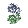
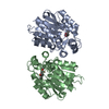
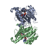
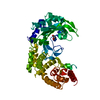
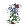
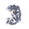
| Title | Native Form 2 E.coli Chaperone Hsp31 | ||||||
 Components Components | Chaperone protein hchA | ||||||
 Keywords Keywords | CHAPERONE / Heat Shock Protein / Putative Catalytic Triad / Flexibility / monoclinic | ||||||
| Function / homology |  Function and homology information Function and homology informationD-lactate dehydratase / response to methylglyoxal / thiolester hydrolase activity / glyoxalase III activity / : / guanine deglycation / protein deglycase / protein deglycase activity / RNA repair / protein repair ...D-lactate dehydratase / response to methylglyoxal / thiolester hydrolase activity / glyoxalase III activity / : / guanine deglycation / protein deglycase / protein deglycase activity / RNA repair / protein repair / Hydrolases; Acting on ester bonds; Thioester hydrolases / Hydrolases; Acting on carbon-nitrogen bonds, other than peptide bonds; In linear amides / response to acidic pH / DNA repair / protein homodimerization activity / zinc ion binding / cytosol / cytoplasm Similarity search - Function | ||||||
| Biological species |  | ||||||
| Method |  X-RAY DIFFRACTION / SYNCHROTRON / MOLECULAR REPLACEMENT / Resolution: 2.71 Å X-RAY DIFFRACTION / SYNCHROTRON / MOLECULAR REPLACEMENT / Resolution: 2.71 Å | ||||||
 Authors Authors | Quigley, P.M. / Korotkov, K. / Baneyx, F. / Hol, W.G.J. | ||||||
 Citation Citation | Journal: Protein Sci. / Year: 2004 Title: A new native EcHsp31 structure suggests a key role of structural flexibility for chaperone function. Authors: Quigley, P.M. / Korotkov, K. / Baneyx, F. / Hol, W.G.J. | ||||||
| History |
|
- Structure visualization
| Structure viewer | Molecule: MolmilJmol/JSmol |
|---|
- Downloads & links
-Download
| PDBx/mmCIF format | 1pv2.cif.gz | 398.5 KB | Display | PDBx/mmCIF format |
|---|---|---|---|---|
| PDB format | pdb1pv2.ent.gz | 329.2 KB | Display | PDB format |
| PDBx/mmJSON format | 1pv2.json.gz | Tree view | PDBx/mmJSON format | |
| Others |  Other downloads Other downloads |
-Validation report
| Arichive directory | https://data.pdbj.org/pub/pdb/validation_reports/pv/1pv2ftp://data.pdbj.org/pub/pdb/validation_reports/pv/1pv2 | HTTPS FTP |
|---|
-Related structure data
| Related structure data |  1n57S S: Starting model for refinement |
|---|---|
| Similar structure data |


-Links
 PDBj
PDBj


- Assembly
| Deposited unit | 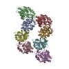
| ||||||||
|---|---|---|---|---|---|---|---|---|---|
| 1 | 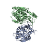
| ||||||||
| 2 | 
| ||||||||
| 3 | 
| ||||||||
| 4 | 
| ||||||||
| Unit cell |
|
-Components
| #1: Protein | Mass: 31226.416 Da / Num. of mol.: 8 Source method: isolated from a genetically manipulated source Source: (gene. exp.) #2: Water | ChemComp-HOH / |  Mass: 18.015 Da / Num. of mol.: 72 / Source method: isolated from a natural source / Formula: H2O Mass: 18.015 Da / Num. of mol.: 72 / Source method: isolated from a natural source / Formula: H2O |
|---|
-Experimental details
-Experiment
| Experiment | Method: X-RAY DIFFRACTION / Number of used crystals: 1 |
|---|
- Sample preparation
| Crystal | Density Matthews: 2.39 Å3/Da / Density % sol: 48.51 % | |||||||||||||||||||||||||||||||||||||||||||||||||||||||||||||||
|---|---|---|---|---|---|---|---|---|---|---|---|---|---|---|---|---|---|---|---|---|---|---|---|---|---|---|---|---|---|---|---|---|---|---|---|---|---|---|---|---|---|---|---|---|---|---|---|---|---|---|---|---|---|---|---|---|---|---|---|---|---|---|---|---|
| Crystal grow | Temperature: 295 K / Method: vapor diffusion, sitting drop / pH: 7.5 Details: 26% PEG 6000,50mM NaCl,100 mM Tris pH 7.5.5G, VAPOR DIFFUSION, SITTING DROP, temperature 22K, temperature 295K | |||||||||||||||||||||||||||||||||||||||||||||||||||||||||||||||
| Crystal grow | *PLUS pH: 7.4 / Method: vapor diffusion | |||||||||||||||||||||||||||||||||||||||||||||||||||||||||||||||
| Components of the solutions | *PLUS
|
-Data collection
| Diffraction | Mean temperature: 100 K |
|---|---|
| Diffraction source | Source: SYNCHROTRON / Site: ALS  / Beamline: 8.2.1 / Wavelength: 1.77 Å / Beamline: 8.2.1 / Wavelength: 1.77 Å |
| Detector | Detector: CCD / Date: Jan 13, 2002 |
| Radiation | Protocol: SINGLE WAVELENGTH / Monochromatic (M) / Laue (L): M / Scattering type: x-ray |
| Radiation wavelength | Wavelength: 1.77 Å / Relative weight: 1 |
| Reflection | Resolution: 2.71→48.8 Å / Num. all: 57067 / Num. obs: 57067 / % possible obs: 100 % / Observed criterion σ(I): 28 / Redundancy: 1.9 % / Rmerge(I) obs: 0.075 / Rsym value: 0.075 / Net I/σ(I): 9.4 |
| Reflection shell | Resolution: 2.7→2.8 Å / Redundancy: 1.8 % / Rmerge(I) obs: 0.339 / Mean I/σ(I) obs: 2 / Num. unique all: 5883 / Rsym value: 0.339 / % possible all: 94.5 |
| Reflection | *PLUS Highest resolution: 2.7 Å / Num. obs: 60105 / % possible obs: 96.1 % |
| Reflection shell | *PLUS % possible obs: 94.1 % / Mean I/σ(I) obs: 2 |
- Processing
| Software |
| ||||||||||||||||||||||||||||||||||||||||||||||||||||||||||||||||||||||
|---|---|---|---|---|---|---|---|---|---|---|---|---|---|---|---|---|---|---|---|---|---|---|---|---|---|---|---|---|---|---|---|---|---|---|---|---|---|---|---|---|---|---|---|---|---|---|---|---|---|---|---|---|---|---|---|---|---|---|---|---|---|---|---|---|---|---|---|---|---|---|---|
| Refinement | Method to determine structure: MOLECULAR REPLACEMENT Starting model: pdb entry 1n57 Resolution: 2.71→48.8 Å / Cor.coef. Fo:Fc: 0.917 / Cor.coef. Fo:Fc free: 0.863 / SU B: 16.066 / SU ML: 0.312 / Cross valid method: THROUGHOUT / ESU R Free: 0.421 Stereochemistry target values: MAXIMUM LIKELIHOOD WITH PHASES
| ||||||||||||||||||||||||||||||||||||||||||||||||||||||||||||||||||||||
| Solvent computation | Ion probe radii: 0.8 Å / Shrinkage radii: 0.8 Å / VDW probe radii: 1.4 Å / Solvent model: BABINET MODEL WITH MASK | ||||||||||||||||||||||||||||||||||||||||||||||||||||||||||||||||||||||
| Displacement parameters | Biso mean: 45.455 Å2
| ||||||||||||||||||||||||||||||||||||||||||||||||||||||||||||||||||||||
| Refinement step | Cycle: LAST / Resolution: 2.71→48.8 Å
| ||||||||||||||||||||||||||||||||||||||||||||||||||||||||||||||||||||||
| Refine LS restraints |
| ||||||||||||||||||||||||||||||||||||||||||||||||||||||||||||||||||||||
| LS refinement shell | Resolution: 2.707→2.777 Å / Total num. of bins used: 20 /
| ||||||||||||||||||||||||||||||||||||||||||||||||||||||||||||||||||||||
| Refinement | *PLUS Highest resolution: 2.7 Å / Lowest resolution: 20 Å / Rfactor Rfree: 0.287 / Rfactor Rwork: 0.222 | ||||||||||||||||||||||||||||||||||||||||||||||||||||||||||||||||||||||
| Solvent computation | *PLUS | ||||||||||||||||||||||||||||||||||||||||||||||||||||||||||||||||||||||
| Displacement parameters | *PLUS | ||||||||||||||||||||||||||||||||||||||||||||||||||||||||||||||||||||||
| Refine LS restraints | *PLUS
|
