 Movie
Movie Controller

[English] 日本語
 Yorodumi
- PDB-1lxc: Crystal Structure of E. Coli Enoyl Reductase-NAD+ with a Bound Ac... -
+ Open data

- Basic information
| Entry | Database: PDB / ID: 1lxc | ||||||
|---|---|---|---|---|---|---|---|
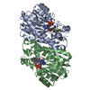
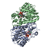
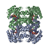
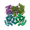
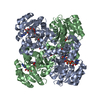
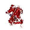
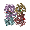
| Title | Crystal Structure of E. Coli Enoyl Reductase-NAD+ with a Bound Acrylamide Inhibitor | ||||||
 Components Components | ENOYL-[ACYL-CARRIER-PROTEIN] REDUCTASE [NADH] | ||||||
 Keywords Keywords | OXIDOREDUCTASE / FABI / ENOYL REDUCTASE | ||||||
| Function / homology |  Function and homology information Function and homology informationenoyl-[acyl-carrier-protein] reductase [NAD(P)H] activity / NADH binding / biotin biosynthetic process / enoyl-[acyl-carrier-protein] reductase (NADH) / fatty acid elongation / enoyl-[acyl-carrier-protein] reductase (NADH) activity / lipid biosynthetic process / catalytic complex / protein homotetramerization / response to antibiotic ...enoyl-[acyl-carrier-protein] reductase [NAD(P)H] activity / NADH binding / biotin biosynthetic process / enoyl-[acyl-carrier-protein] reductase (NADH) / fatty acid elongation / enoyl-[acyl-carrier-protein] reductase (NADH) activity / lipid biosynthetic process / catalytic complex / protein homotetramerization / response to antibiotic / protein-containing complex / membrane / identical protein binding / cytosol Similarity search - Function | ||||||
| Biological species |  | ||||||
| Method |  X-RAY DIFFRACTION / SYNCHROTRON / MOLECULAR REPLACEMENT / Resolution: 2.4 Å X-RAY DIFFRACTION / SYNCHROTRON / MOLECULAR REPLACEMENT / Resolution: 2.4 Å | ||||||
 Authors Authors | Miller, W.H. / Seefeld, M.A. / Newlander, K.A. / Uzinskas, I.N. / Burgess, W.J. / Heerding, D.A. / Yuan, C.C.K. / Head, M.S. / Payne, D.J. / Rittenhouse, S.F. ...Miller, W.H. / Seefeld, M.A. / Newlander, K.A. / Uzinskas, I.N. / Burgess, W.J. / Heerding, D.A. / Yuan, C.C.K. / Head, M.S. / Payne, D.J. / Rittenhouse, S.F. / Moore, T.D. / Pearson, S.C. / Dewolf, V. / Berry, W.E. / Keller, P.M. / Polizzi, B.J. / Qiu, X. / Janson, C.A. / Huffman, W.F. | ||||||
 Citation Citation | Journal: J.Med.Chem. / Year: 2002 Title: Discovery of aminopyridine-based inhibitors of bacterial enoyl-ACP reductase (FabI). Authors: Miller, W.H. / Seefeld, M.A. / Newlander, K.A. / Uzinskas, I.N. / Burgess, W.J. / Heerding, D.A. / Yuan, C.C. / Head, M.S. / Payne, D.J. / Rittenhouse, S.F. / Moore, T.D. / Pearson, S.C. / ...Authors: Miller, W.H. / Seefeld, M.A. / Newlander, K.A. / Uzinskas, I.N. / Burgess, W.J. / Heerding, D.A. / Yuan, C.C. / Head, M.S. / Payne, D.J. / Rittenhouse, S.F. / Moore, T.D. / Pearson, S.C. / Berry, V. / DeWolf Jr., W.E. / Keller, P.M. / Polizzi, B.J. / Qiu, X. / Janson, C.A. / Huffman, W.F. | ||||||
| History |
|
- Structure visualization
| Structure viewer | Molecule: MolmilJmol/JSmol |
|---|
- Downloads & links
-Download
| PDBx/mmCIF format | 1lxc.cif.gz | 114.4 KB | Display | PDBx/mmCIF format |
|---|---|---|---|---|
| PDB format | pdb1lxc.ent.gz | 88.8 KB | Display | PDB format |
| PDBx/mmJSON format | 1lxc.json.gz | Tree view | PDBx/mmJSON format | |
| Others |  Other downloads Other downloads |
-Validation report
| Arichive directory | https://data.pdbj.org/pub/pdb/validation_reports/lx/1lxcftp://data.pdbj.org/pub/pdb/validation_reports/lx/1lxc | HTTPS FTP |
|---|
-Related structure data
| Related structure data |  1lx6C 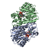 1c14S S: Starting model for refinement C: citing same article ( |
|---|---|
| Similar structure data |


-Links
 PDBj
PDBj

- Assembly
| Deposited unit | 
| ||||||||
|---|---|---|---|---|---|---|---|---|---|
| 1 | 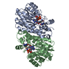
| ||||||||
| Unit cell |
| ||||||||
| Details | THE ASYMMETRIC UNIT CAONTAINS A DIMER OF THE TERNARY COMPLEX |
-Components
| #1: Protein | Mass: 27892.926 Da / Num. of mol.: 2 / Source method: isolated from a natural source / Source: (natural) References: UniProt: P29132, UniProt: P0AEK4*PLUS, enoyl-[acyl-carrier-protein] reductase (NADH) #2: Chemical |   Mass: 663.425 Da / Num. of mol.: 2 / Source method: obtained synthetically / Formula: C21H27N7O14P2 / Comment: NAD*YM Mass: 663.425 Da / Num. of mol.: 2 / Source method: obtained synthetically / Formula: C21H27N7O14P2 / Comment: NAD*YM#3: Chemical |   Mass: 320.388 Da / Num. of mol.: 2 / Source method: obtained synthetically / Formula: C19H20N4O Mass: 320.388 Da / Num. of mol.: 2 / Source method: obtained synthetically / Formula: C19H20N4O#4: Water | ChemComp-HOH / |  Mass: 18.015 Da / Num. of mol.: 300 / Source method: isolated from a natural source / Formula: H2O Mass: 18.015 Da / Num. of mol.: 300 / Source method: isolated from a natural source / Formula: H2O |
|---|
-Experimental details
-Experiment
| Experiment | Method: X-RAY DIFFRACTION / Number of used crystals: 1 |
|---|
- Sample preparation
| Crystal | Density Matthews: 2.67 Å3/Da / Density % sol: 53.96 % | ||||||||||||||||||||||||||||||||||||
|---|---|---|---|---|---|---|---|---|---|---|---|---|---|---|---|---|---|---|---|---|---|---|---|---|---|---|---|---|---|---|---|---|---|---|---|---|---|
| Crystal grow | Temperature: 298 K / Method: vapor diffusion, sitting drop / pH: 7.5 Details: 0.1 M HEPES, 2M(NH4)2SO4,5% PEG400, pH 7.5, VAPOR DIFFUSION, SITTING DROP, temperature 298K | ||||||||||||||||||||||||||||||||||||
| Crystal grow | *PLUS Details: Qiu, X., (1999) Protein Sci., 8, 2529. | ||||||||||||||||||||||||||||||||||||
| Components of the solutions | *PLUS
|
-Data collection
| Diffraction | Mean temperature: 100 K |
|---|---|
| Diffraction source | Source: SYNCHROTRON / Site: NSLS  / Beamline: X12C / Wavelength: 1 Å / Beamline: X12C / Wavelength: 1 Å |
| Detector | Type: BRANDEIS / Detector: CCD / Date: Nov 22, 1999 |
| Radiation | Monochromator: Si / Protocol: SINGLE WAVELENGTH / Monochromatic (M) / Laue (L): M / Scattering type: x-ray |
| Radiation wavelength | Wavelength: 1 Å / Relative weight: 1 |
| Reflection | Resolution: 2.4→20 Å / Num. all: 25117 / Num. obs: 20621 / % possible obs: 82.1 % / Observed criterion σ(F): 2 / Observed criterion σ(I): 2 / Redundancy: 4.5 % / Biso Wilson estimate: 22.7 Å2 / Rmerge(I) obs: 0.053 / Net I/σ(I): 22.5 |
| Reflection shell | Resolution: 2.4→2.44 Å / Rmerge(I) obs: 0.098 / Mean I/σ(I) obs: 6.9 / % possible all: 43.2 |
| Reflection | *PLUS Num. measured all: 91795 / Rmerge(I) obs: 0.053 |
| Reflection shell | *PLUS % possible obs: 43.2 % / Rmerge(I) obs: 0.098 |
- Processing
| Software |
| ||||||||||||||||||||||||||||||||||||||||||||||||||||||||||||
|---|---|---|---|---|---|---|---|---|---|---|---|---|---|---|---|---|---|---|---|---|---|---|---|---|---|---|---|---|---|---|---|---|---|---|---|---|---|---|---|---|---|---|---|---|---|---|---|---|---|---|---|---|---|---|---|---|---|---|---|---|---|
| Refinement | Method to determine structure: MOLECULAR REPLACEMENT Starting model: 1C14 Resolution: 2.4→20 Å / Rfactor Rfree error: 0.009 / Isotropic thermal model: RESTRAINED / Cross valid method: THROUGHOUT / σ(F): 2 / σ(I): 2 / Stereochemistry target values: Engh & Huber
| ||||||||||||||||||||||||||||||||||||||||||||||||||||||||||||
| Displacement parameters | Biso mean: 26.3 Å2
| ||||||||||||||||||||||||||||||||||||||||||||||||||||||||||||
| Refine analyze |
| ||||||||||||||||||||||||||||||||||||||||||||||||||||||||||||
| Refinement step | Cycle: LAST / Resolution: 2.4→20 Å
| ||||||||||||||||||||||||||||||||||||||||||||||||||||||||||||
| Refine LS restraints |
| ||||||||||||||||||||||||||||||||||||||||||||||||||||||||||||
| LS refinement shell | Resolution: 2.4→2.44 Å / Rfactor Rfree error: 0.03 / Total num. of bins used: 6
| ||||||||||||||||||||||||||||||||||||||||||||||||||||||||||||
| Xplor file |
| ||||||||||||||||||||||||||||||||||||||||||||||||||||||||||||
| Refinement | *PLUS Lowest resolution: 20 Å / σ(I): 0 / Rfactor Rfree: 0.253 / Rfactor Rwork: 0.199 | ||||||||||||||||||||||||||||||||||||||||||||||||||||||||||||
| Solvent computation | *PLUS | ||||||||||||||||||||||||||||||||||||||||||||||||||||||||||||
| Displacement parameters | *PLUS | ||||||||||||||||||||||||||||||||||||||||||||||||||||||||||||
| Refine LS restraints | *PLUS
| ||||||||||||||||||||||||||||||||||||||||||||||||||||||||||||
| LS refinement shell | *PLUS Rfactor Rfree: 0.2555 / Rfactor Rwork: 0.215 |
